• 文献检索
  • 文档翻译
  • 深度研究
  • 学术资讯
  • Suppr Zotero 插件Zotero 插件
  • 邀请有礼
  • 套餐&价格
  • 历史记录
应用&插件
Suppr Zotero 插件Zotero 插件浏览器插件Mac 客户端Windows 客户端微信小程序
定价
高级版会员购买积分包购买API积分包
服务
文献检索文档翻译深度研究API 文档MCP 服务
关于我们
关于 Suppr公司介绍联系我们用户协议隐私条款
关注我们

Suppr 超能文献

核心技术专利:CN118964589B侵权必究
粤ICP备2023148730 号-1Suppr @ 2026

文献检索

告别复杂PubMed语法,用中文像聊天一样搜索,搜遍4000万医学文献。AI智能推荐,让科研检索更轻松。

立即免费搜索

文件翻译

保留排版,准确专业,支持PDF/Word/PPT等文件格式,支持 12+语言互译。

免费翻译文档

深度研究

AI帮你快速写综述,25分钟生成高质量综述,智能提取关键信息,辅助科研写作。

立即免费体验

室管膜瘤的影像学检查:磁共振成像(MRI)和计算机断层扫描(CT)

Imaging of ependymomas: MRI and CT.

作者信息

Yuh E L, Barkovich A J, Gupta N

机构信息

Neuroradiology Section, Department of Radiology, University of California, San Francisco, San Francisco, USA.

出版信息

Childs Nerv Syst. 2009 Oct;25(10):1203-13. doi: 10.1007/s00381-009-0878-7. Epub 2009 Apr 10.

DOI:10.1007/s00381-009-0878-7
PMID:19360419
原文链接:https://pmc.ncbi.nlm.nih.gov/articles/PMC2744772/
Abstract

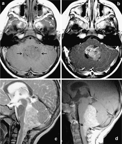
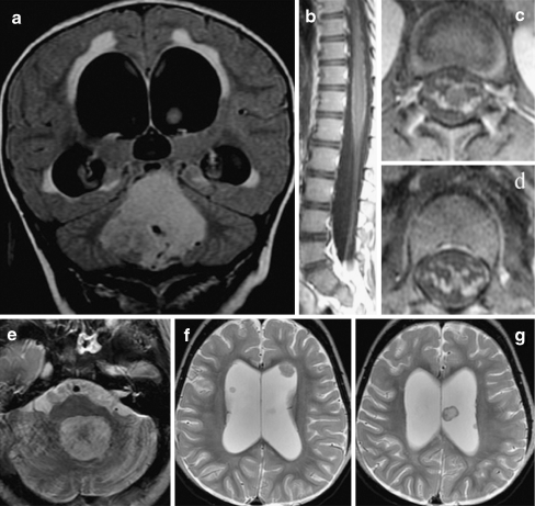
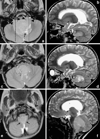
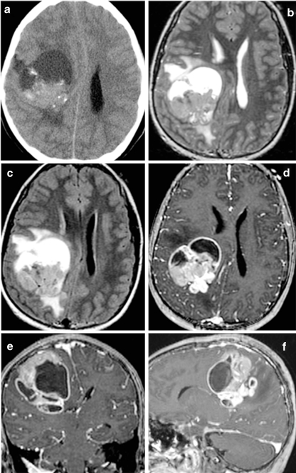
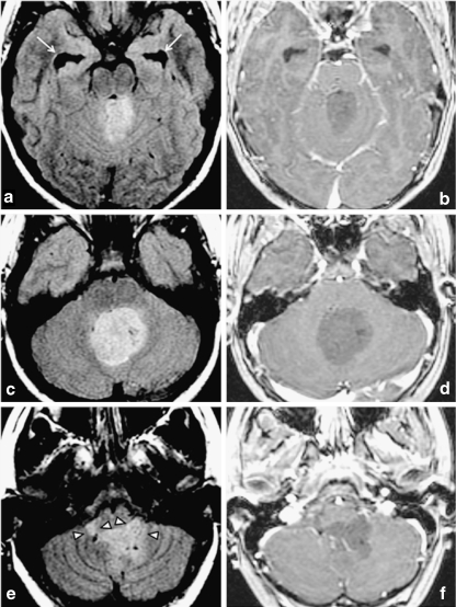
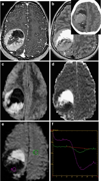
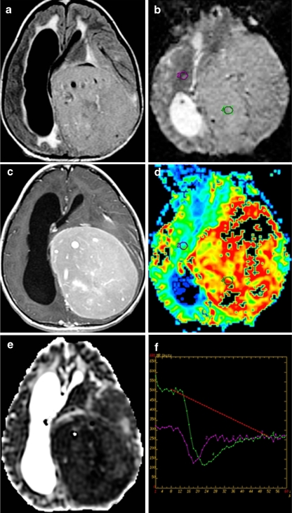
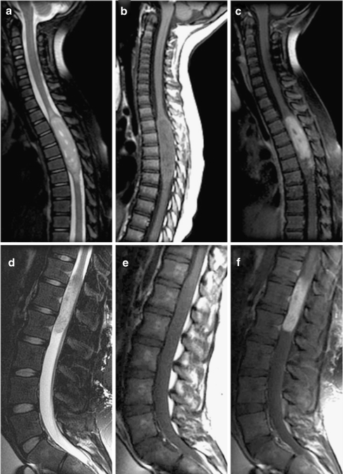
The imaging features of intracranial and spinal ependymoma are reviewed with an emphasis on conventional magnetic resonance imaging (MRI), perfusion MRI and proton magnetic resonance spectroscopy, and computed tomography. Imaging manifestations of leptomeningeal dissemination of disease are described. Finally, salient imaging features obtained in the postoperative period to evaluate completeness of surgical resection, and thereafter for long-term surveillance for disease recurrence, are reviewed.

摘要

本文回顾了颅内和脊髓室管膜瘤的影像学特征,重点介绍了传统磁共振成像(MRI)、灌注MRI、质子磁共振波谱以及计算机断层扫描。描述了疾病软脑膜播散的影像学表现。最后,回顾了术后用于评估手术切除完整性以及随后长期监测疾病复发的显著影像学特征。

https://cdn.ncbi.nlm.nih.gov/pmc/blobs/005d/2744772/58aa005d6f8c/381_2009_878_Fig9_HTML.jpg
https://cdn.ncbi.nlm.nih.gov/pmc/blobs/005d/2744772/823181caf07c/381_2009_878_Fig1_HTML.jpg
https://cdn.ncbi.nlm.nih.gov/pmc/blobs/005d/2744772/2383a3e37e2a/381_2009_878_Fig2_HTML.jpg
https://cdn.ncbi.nlm.nih.gov/pmc/blobs/005d/2744772/5d8f22bd8bce/381_2009_878_Fig3_HTML.jpg
https://cdn.ncbi.nlm.nih.gov/pmc/blobs/005d/2744772/c8930a8d4228/381_2009_878_Fig4_HTML.jpg
https://cdn.ncbi.nlm.nih.gov/pmc/blobs/005d/2744772/60476f277caf/381_2009_878_Fig5_HTML.jpg
https://cdn.ncbi.nlm.nih.gov/pmc/blobs/005d/2744772/a2f6a1c9276a/381_2009_878_Fig6_HTML.jpg
https://cdn.ncbi.nlm.nih.gov/pmc/blobs/005d/2744772/804539bdb890/381_2009_878_Fig7_HTML.jpg
https://cdn.ncbi.nlm.nih.gov/pmc/blobs/005d/2744772/86f50a32d56e/381_2009_878_Fig8_HTML.jpg
https://cdn.ncbi.nlm.nih.gov/pmc/blobs/005d/2744772/58aa005d6f8c/381_2009_878_Fig9_HTML.jpg

相似文献

1
Imaging of ependymomas: MRI and CT.室管膜瘤的影像学检查:磁共振成像(MRI)和计算机断层扫描(CT)
Childs Nerv Syst. 2009 Oct;25(10):1203-13. doi: 10.1007/s00381-009-0878-7. Epub 2009 Apr 10.
2
Spinal extramedullary anaplastic ependymoma with spinal and intracranial metastases.伴有脊柱和颅内转移的脊髓髓外间变性室管膜瘤
J Neurooncol. 2006 Aug;79(1):57-9. doi: 10.1007/s11060-005-9114-9. Epub 2006 Apr 14.
3
Intracranial ependymomas: The role of advanced neuroimaging in diagnosis and management.颅内室管膜瘤:高级神经影像学在诊断和治疗中的作用。
Neuroradiol J. 2021 Apr;34(2):80-92. doi: 10.1177/1971400921990770. Epub 2021 Feb 1.
4
A case of cervicomedullary junction tanycytic ependymoma associated with marked cyst formation.一例伴有明显囊肿形成的颈髓交界区室管膜瘤。
Brain Tumor Pathol. 2005;22(1):29-33. doi: 10.1007/s10014-005-0174-5.
5
Multiple intradural-extramedullary spinal ependymomas including tumors with different histological features.多发髓内硬膜外脊髓室管膜瘤,包括具有不同组织学特征的肿瘤。
Eur Spine J. 2017 May;26(Suppl 1):222-224. doi: 10.1007/s00586-017-5055-1. Epub 2017 Mar 30.
6
Comparison of Conventional, Diffusion, and Perfusion MRI Between Low-Grade and Anaplastic Extraventricular Ependymoma.低级别与间变性室管膜瘤的常规弥散与灌注 MRI 对比。
AJR Am J Roentgenol. 2020 Oct;215(4):978-984. doi: 10.2214/AJR.20.22764. Epub 2020 Aug 18.
7
Myxopapillary Ependymoma.黏液乳头型室管膜瘤
Radiographics. 2019 Mar-Apr;39(2):467. doi: 10.1148/rg.2019184014.
8
Natural Course of Myxopapillary Ependymoma: Unusual Case Report and Review of Literature.黏液乳头型室管膜瘤的自然病程:罕见病例报告及文献综述
World Neurosurg. 2019 Jan;121:239-242. doi: 10.1016/j.wneu.2018.10.028. Epub 2018 Oct 12.
9
[Lumbosciatica revealing intramedullary ependymoma].[以腰骶部坐骨神经痛为表现的髓内室管膜瘤]
Pan Afr Med J. 2017 Mar 24;26:171. doi: 10.11604/pamj.2017.26.171.11633. eCollection 2017.
10
Differential involvement of protein 4.1 family members DAL-1 and NF2 in intracranial and intraspinal ependymomas.蛋白质4.1家族成员DAL-1和NF2在颅内和脊髓室管膜瘤中的不同作用。
Mod Pathol. 2002 May;15(5):526-31. doi: 10.1038/modpathol.3880558.

引用本文的文献

1
Correlation Analysis of Imaging and Pathological Features of Ependymomas.室管膜瘤影像学与病理学特征的相关性分析
Int J Gen Med. 2025 Jul 21;18:4001-4009. doi: 10.2147/IJGM.S528992. eCollection 2025.
2
Spinal lesions: a comprehensive radiologic overview.脊柱病变:全面的放射学概述
Front Radiol. 2025 Jun 27;5:1577840. doi: 10.3389/fradi.2025.1577840. eCollection 2025.
3
Dynamic Magnetic Resonance Imaging Changes in Spinal Ependymomas and Their Impact on Surgical Planning.脊髓室管膜瘤的动态磁共振成像变化及其对手术规划的影响

本文引用的文献

1
Cerebrospinal fluid cytology in patients with ependymoma: a bi-institutional retrospective study.室管膜瘤患者的脑脊液细胞学检查:一项双机构回顾性研究。
Cancer. 2008 Oct 25;114(5):307-14. doi: 10.1002/cncr.23799.
2
Radiation risk to children from computed tomography.计算机断层扫描对儿童的辐射风险。
Pediatrics. 2007 Sep;120(3):677-82. doi: 10.1542/peds.2007-1910.
3
Apparent diffusion coefficients for differentiation of cerebellar tumors in children.表观扩散系数在儿童小脑肿瘤鉴别诊断中的应用
Neurol Med Chir (Tokyo). 2025 Jul 15;65(7):310-318. doi: 10.2176/jns-nmc.2024-0328. Epub 2025 May 29.
4
Preoperative MR - based model for predicting prognosis in patients with intracranial extraventricular ependymoma.基于术前磁共振成像的模型预测颅内室管膜外瘤患者的预后
Eur J Radiol Open. 2025 Apr 8;14:100650. doi: 10.1016/j.ejro.2025.100650. eCollection 2025 Jun.
5
Beyond the expected: a supratentorial ependymoma imitating a meningioma.超乎预期:一例幕上室管膜瘤酷似脑膜瘤
Childs Nerv Syst. 2025 Mar 26;41(1):141. doi: 10.1007/s00381-025-06803-7.
6
Progression and diagnostic challenges of desmoplastic infantile ganglioglioma in a non-infant: a case report with 5-year follow-up.非婴儿型促结缔组织增生性婴儿型神经节胶质瘤的进展及诊断挑战:一例5年随访病例报告
Front Oncol. 2025 Feb 10;15:1411213. doi: 10.3389/fonc.2025.1411213. eCollection 2025.
7
Intracranial ependymoma: A retrospective analysis of clinical features, treatment modalities, and long-term outcome.颅内室管膜瘤:临床特征、治疗方式及长期预后的回顾性分析
Aging Med (Milton). 2024 Dec 11;7(6):679-688. doi: 10.1002/agm2.12378. eCollection 2024 Dec.
8
Machine learning-based nomogram for distinguishing between supratentorial extraventricular ependymoma and supratentorial glioblastoma.基于机器学习的列线图用于鉴别幕上脑室外室管膜瘤和幕上胶质母细胞瘤
Front Oncol. 2024 Sep 9;14:1443913. doi: 10.3389/fonc.2024.1443913. eCollection 2024.
9
Supratentorial and Infratentorial Ependymoma.幕上和幕下室管膜瘤。
Adv Tech Stand Neurosurg. 2024;53:93-118. doi: 10.1007/978-3-031-67077-0_7.
10
Ventricular system-unrelated cerebellar ependymoma: A case report.脑室系统无关的小脑室管膜瘤:一例报告。
World J Clin Cases. 2024 Sep 6;12(25):5814-5820. doi: 10.12998/wjcc.v12.i25.5814.
AJNR Am J Neuroradiol. 2006 Jun-Jul;27(6):1362-9.
4
Intracranial ependymoma.颅内室管膜瘤
Neurosurg Focus. 2002 Sep 15;13(3):e4. doi: 10.3171/foc.2002.13.3.5.
5
Best cases from the AFIP: supratentorial ependymoma.武装部队病理研究所的最佳病例:幕上室管膜瘤。
Radiographics. 2005 Mar-Apr;25(2):486-90. doi: 10.1148/rg.252045095.
6
Intraventricular tanycytic ependymoma: case report and review of the literature.脑室内室管膜下巨细胞型室管膜瘤:病例报告及文献复习
J Neurooncol. 2005 Jan;71(2):189-93. doi: 10.1007/s11060-004-1371-5.
7
From the archives of the AFIP. Cerebral intraventricular neoplasms: radiologic-pathologic correlation.源自武装部队病理研究所档案。脑室内肿瘤:放射学与病理学对照。
Radiographics. 2002 Nov-Dec;22(6):1473-505. doi: 10.1148/rg.226025118.
8
Pattern of recurrence in children with midline posterior fossa malignant neoplasms.
Pediatr Radiol. 2000 Feb;30(2):90-3. doi: 10.1007/s002470050022.
9
MRI features of intracranial and spinal ependymomas.颅内和脊髓室管膜瘤的磁共振成像特征
Pediatr Neurosurg. 1998 Feb;28(2):97-105. doi: 10.1159/000028630.
10
Clinical manifestations of childhood ependymoma: a multitude of syndromes.儿童室管膜瘤的临床表现:多种综合征
Pediatr Neurosurg. 1998 Jan;28(1):49-55. doi: 10.1159/000028619.