Rulong S, Aguas A P, da Silva P P, Silva M T
Section of Membrane Biology, National Cancer Institute-Frederick Cancer Research and Development Center, Maryland 21702-1201.
Infect Immun. 1991 Nov;59(11):3895-902. doi: 10.1128/iai.59.11.3895-3902.1991.
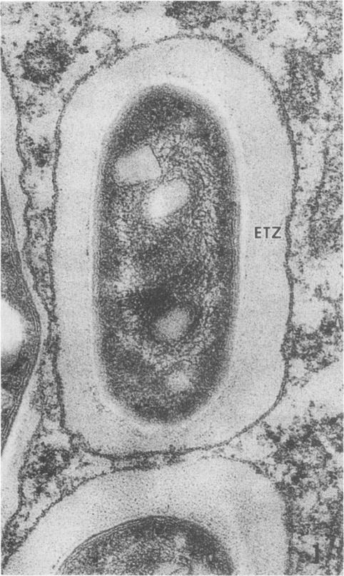
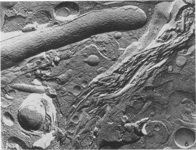
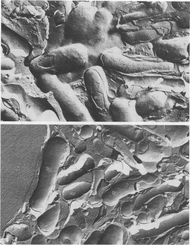
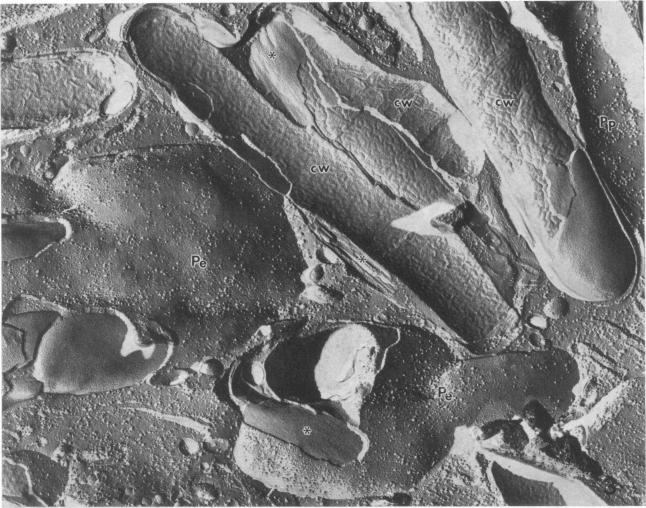
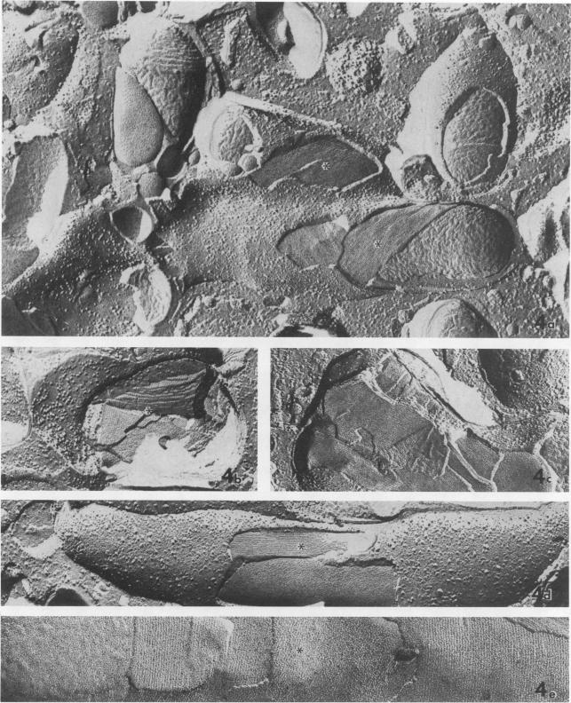
We used freeze fracture electron microscopy to study the fine structure of Mycobacterium avium inside phagosomes of murine macrophages. M. avium-susceptible C57BL/6 mice were infected with M. avium by intraperitoneal inoculation of 10(8) viable bacilli. We studied the microanatomy of the mycobacteria in 3-month infections of mice, a situation in which bacillary multiplication is extensive. In these samples, freeze fracture revealed that intraphagosomal bacilli were surrounded by a multilamellar coat that was apposed to the cell wall. In thin sections, in contrast, the area corresponding to the coat showed no substructure and was electron transparent (the so-called electron-transparent zone that has been previously reported by others). The multiple lamellae resembled an onionlike assembly that was inserted in between the mycobacterial wall outer surface and the phagosomal membrane. Each lamella of the M. avium coat was made up of parallel straight fibrils with a width of 5 nm. A variable number of lamellae, sometimes up to 10 or more elements, coated individual bacilli. The multilamellar coat was absent around both extracellular M. avium and intramacrophagic M. avium after short-term (45-min) inoculation of mice. The supramolecular organization of the M. avium lamellar coat as viewed here by freeze fracture is similar to that of purified mycoside C (P. Draper, J. Gen. Microbiol. 83:431-433, 1974; K.-S. Kim, M.R.J. Salton, and L. Barksdale, J. Bacteriol. 125:739-743, 1976), a mycobacterial component currently known as glycopeptidolipid (W.W. Barrow and P.J. Brennan, J. Bacteriol. 150:381-384, 1982). We conclude that M. avium bacilli growing in macrophages are surrounded by multilamellar capsulelike structures that contain glycopeptidolipid molecules.
我们使用冷冻断裂电子显微镜来研究鼠巨噬细胞吞噬体中鸟分枝杆菌的精细结构。对鸟分枝杆菌易感的C57BL/6小鼠通过腹腔接种10⁸ 个活的杆菌来感染鸟分枝杆菌。我们研究了小鼠感染3个月时分枝杆菌的微观解剖结构,此时杆菌大量繁殖。在这些样本中,冷冻断裂显示吞噬体内的杆菌被一层与细胞壁相邻的多层膜包被所包围。相比之下,在薄切片中,对应于该包被的区域没有亚结构且电子透明(即先前其他人报道过的所谓电子透明区)。多层膜类似于插入分枝杆菌细胞壁外表面和吞噬体膜之间的洋葱状结构。鸟分枝杆菌包被的每一层由宽度为5纳米的平行直纤维组成。不同数量的层,有时多达10层或更多,包裹着单个杆菌。在给小鼠短期(45分钟)接种后,胞外鸟分枝杆菌和巨噬细胞内的鸟分枝杆菌周围均不存在多层包被。此处通过冷冻断裂观察到的鸟分枝杆菌层状包被的超分子组织与纯化的霉菌糖苷C(P. Draper,《普通微生物学杂志》83:431 - 433,1974;K.-S. Kim,M.R.J. Salton和L. Barksdale,《细菌学杂志》125:739 - 743,1976)相似,霉菌糖苷C是一种目前已知为糖肽脂的分枝杆菌成分(W.W. Barrow和P.J. Brennan,《细菌学杂志》150:381 - 384,1982)。我们得出结论,在巨噬细胞中生长的鸟分枝杆菌杆菌被含有糖肽脂分子的多层囊状结构所包围。