Brun Caroline C, Leporé Natasha, Pennec Xavier, Lee Agatha D, Barysheva Marina, Madsen Sarah K, Avedissian Christina, Chou Yi-Yu, de Zubicaray Greig I, McMahon Katie L, Wright Margaret J, Toga Arthur W, Thompson Paul M
Laboratory of Neuro Imaging, Department of Neurology, UCLA School of Medicine, 635 Charles Young Drive South Suite 225, Los Angeles, CA 90095-7334, USA.
Neuroimage. 2009 Oct 15;48(1):37-49. doi: 10.1016/j.neuroimage.2009.05.022. Epub 2009 May 14.
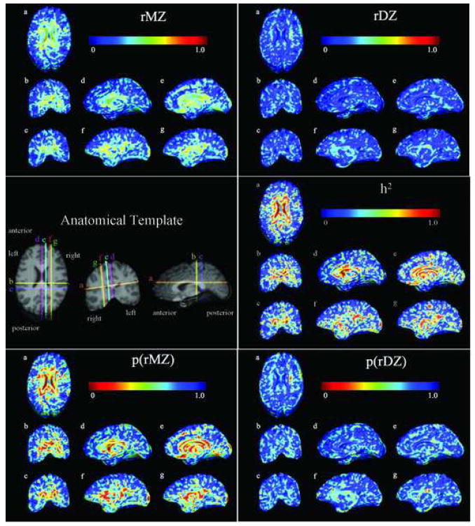
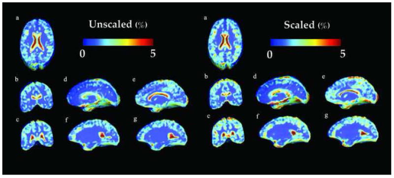
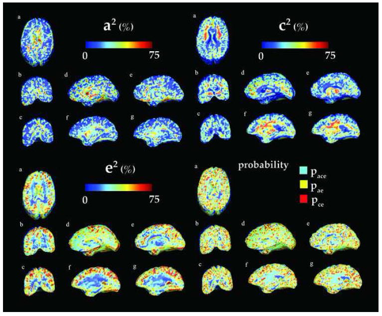
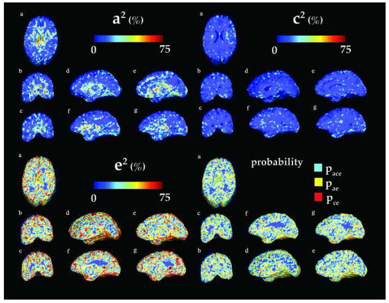
Genetic and environmental factors influence brain structure and function profoundly. The search for heritable anatomical features and their influencing genes would be accelerated with detailed 3D maps showing the degree to which brain morphometry is genetically determined. As part of an MRI study that will scan 1150 twins, we applied Tensor-Based Morphometry to compute morphometric differences in 23 pairs of identical twins and 23 pairs of same-sex fraternal twins (mean age: 23.8+/-1.8 SD years). All 92 twins' 3D brain MRI scans were nonlinearly registered to a common space using a Riemannian fluid-based warping approach to compute volumetric differences across subjects. A multi-template method was used to improve volume quantification. Vector fields driving each subject's anatomy onto the common template were analyzed to create maps of local volumetric excesses and deficits relative to the standard template. Using a new structural equation modeling method, we computed the voxelwise proportion of variance in volumes attributable to additive (A) or dominant (D) genetic factors versus shared environmental (C) or unique environmental factors (E). The method was also applied to various anatomical regions of interest (ROIs). As hypothesized, the overall volumes of the brain, basal ganglia, thalamus, and each lobe were under strong genetic control; local white matter volumes were mostly controlled by common environment. After adjusting for individual differences in overall brain scale, genetic influences were still relatively high in the corpus callosum and in early-maturing brain regions such as the occipital lobes, while environmental influences were greater in frontal brain regions that have a more protracted maturational time-course.
遗传和环境因素对大脑结构和功能有着深远影响。借助详细的三维地图来显示大脑形态测量在多大程度上由基因决定,将加速对可遗传解剖特征及其影响基因的研究。作为一项将对1150对双胞胎进行扫描的磁共振成像(MRI)研究的一部分,我们应用基于张量的形态测量法来计算23对同卵双胞胎和23对同性异卵双胞胎(平均年龄:23.8±1.8标准差岁)之间的形态测量差异。使用基于黎曼流体的变形方法,将所有92名双胞胎的三维脑部MRI扫描非线性配准到一个公共空间,以计算不同受试者之间的体积差异。采用多模板方法来改进体积量化。分析将每个受试者的解剖结构映射到公共模板上的向量场,以创建相对于标准模板的局部体积过剩和不足的图谱。使用一种新的结构方程建模方法,我们计算了体积中可归因于加性(A)或显性(D)遗传因素与共享环境(C)或独特环境因素(E)的体素方差比例。该方法还应用于各个感兴趣的解剖区域(ROI)。正如所假设的,大脑、基底神经节、丘脑以及每个脑叶的总体积受到强大的基因控制;局部白质体积大多由共同环境控制。在调整了全脑尺度的个体差异后,胼胝体和枕叶等早熟脑区的遗传影响仍然相对较高,而额叶脑区的环境影响更大,因为额叶脑区的成熟时间进程更为漫长。