Huang Bo, Bates Mark, Zhuang Xiaowei
Howard Hughes Medical Institute, Harvard University, Cambridge, MA 02138, USA.
Annu Rev Biochem. 2009;78:993-1016. doi: 10.1146/annurev.biochem.77.061906.092014.
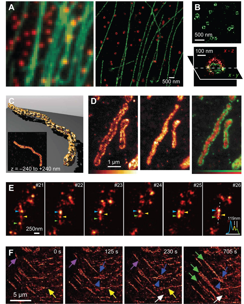
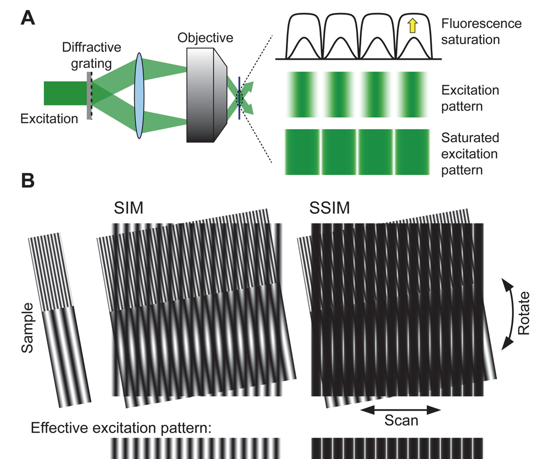
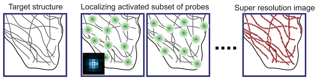
Achieving a spatial resolution that is not limited by the diffraction of light, recent developments of super-resolution fluorescence microscopy techniques allow the observation of many biological structures not resolvable in conventional fluorescence microscopy. New advances in these techniques now give them the ability to image three-dimensional (3D) structures, measure interactions by multicolor colocalization, and record dynamic processes in living cells at the nanometer scale. It is anticipated that super-resolution fluorescence microscopy will become a widely used tool for cell and tissue imaging to provide previously unobserved details of biological structures and processes.
由于实现了不受光衍射限制的空间分辨率,超分辨率荧光显微镜技术的最新进展使得观察许多在传统荧光显微镜下无法分辨的生物结构成为可能。这些技术的新进展使其能够对三维(3D)结构进行成像,通过多色共定位测量相互作用,并在纳米尺度上记录活细胞中的动态过程。预计超分辨率荧光显微镜将成为细胞和组织成像中广泛使用的工具,以提供生物结构和过程以前未被观察到的细节。