Trias Elman L, Hassantoufighi Arash, Prince Gregory A, Eichelberger Maryna C
Children's National Medical Center, Washington, DC 20010, USA.
BMC Pulm Med. 2009 Jun 10;9:28. doi: 10.1186/1471-2466-9-28.
Increased respiratory rate (tachypnea) is frequently observed as a clinical sign of influenza pneumonia in pediatric patients admitted to the hospital. We previously demonstrated that influenza infection of adult cotton rats (Sigmodon hispidus) also results in tachypnea and wanted to establish whether this clinical sign was observed in infected infant cotton rats. We hypothesized that age-dependent differences in lung mechanics result in differences in ventilatory characteristics following influenza infection.
Lung tidal volume, dynamic elastance, resistance, and pleural pressure were measured in a resistance and compliance system on mechanically-ventilated anesthestized young (14-28 day old) and adult (6-12 week old) cotton rats. Animals at the same age were infected with influenza virus, and breathing rates and other respiratory measurements were recorded using a whole body flow plethysmograph.
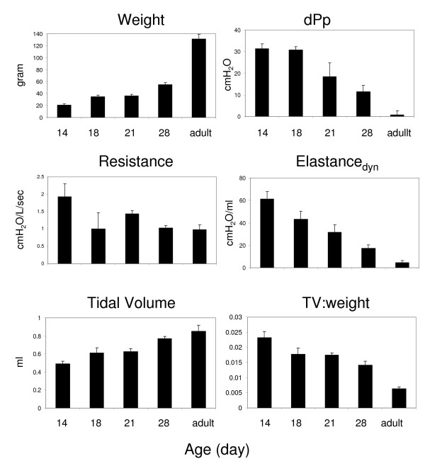
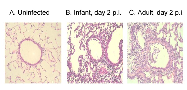
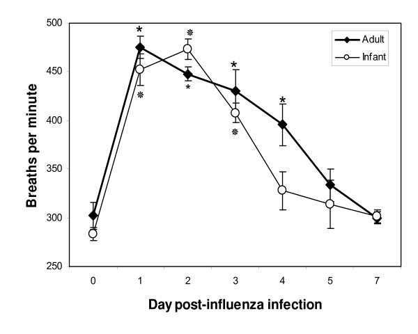
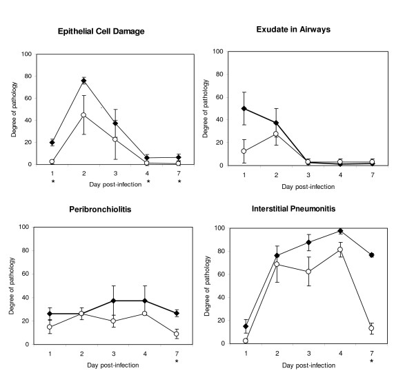
Adult cotton rats had significantly greater tidal volume (TV), and lower resistance and elastance than young animals. To evaluate the impact of this increased lung capacity and stiffening on respiratory disease, young and adult animals were infected intra-nasally with influenza A/Wuhan/359/95. Both age groups had increased respiratory rate and enhanced pause (Penh) during infection, suggesting lower airway obstruction. However, in spite of significant tachypnea, the infant (unlike the adult) cotton rats maintained the same tidal volume, resulting in an increased minute volume. In addition, the parameters that contribute to Penh were different: while relaxation time between breaths and time of expiration was decreased in both age groups, a disproportionate increase in peak inspiratory and expiratory flow contributed to the increase in Penh in infant animals.
While respiratory rate is increased in both adult and infant influenza-infected cotton rats, the volume of air exchanged per minute (minute volume) is increased in the infant animals only. This is likely to be a consequence of greater lung elastance in the very young animals. This model replicates many respiratory features of humans and consequently may be a useful tool to investigate new strategies to treat respiratory disease in influenza-infected infants.
呼吸频率增加(呼吸急促)是住院儿科患者流感肺炎的常见临床症状。我们之前证明,成年棉鼠(棉鼠属)感染流感也会导致呼吸急促,我们想确定在受感染的幼年棉鼠中是否也会出现这种临床症状。我们假设肺力学的年龄依赖性差异会导致流感感染后通气特征的差异。
在机械通气的麻醉幼年(14 - 28日龄)和成年(6 - 12周龄)棉鼠的阻力和顺应性系统中测量肺潮气量、动态弹性、阻力和胸膜压力。相同年龄的动物感染流感病毒,并使用全身血流体积描记器记录呼吸频率和其他呼吸测量值。
成年棉鼠的潮气量(TV)明显大于幼年动物,且阻力和弹性较低。为了评估这种肺容量增加和僵硬对呼吸道疾病的影响,幼年和成年动物经鼻感染甲型流感病毒A/武汉/359/95。两个年龄组在感染期间呼吸频率均增加且增强暂停(Penh),提示存在下呼吸道阻塞。然而,尽管有明显的呼吸急促,幼年(与成年不同)棉鼠的潮气量保持不变,导致每分通气量增加。此外,导致Penh的参数不同:虽然两个年龄组的呼吸间隔时间和呼气时间均减少,但幼年动物中吸气和呼气峰值流量的不成比例增加导致了Penh的增加。
虽然成年和幼年感染流感的棉鼠呼吸频率均增加,但仅幼年动物每分钟交换的空气量(每分通气量)增加。这可能是由于幼年动物肺弹性更大所致。该模型复制了人类的许多呼吸特征,因此可能是研究治疗流感感染婴儿呼吸道疾病新策略的有用工具。