Wilder-Smith Petra, Lee Kenneth, Guo Shuguang, Zhang Jun, Osann Kathryn, Chen Zhongping, Messadi Diana
Beckman Laser Institute, University of California, Irvine, California 92612, USA.
Lasers Surg Med. 2009 Jul;41(5):353-7. doi: 10.1002/lsm.20773.
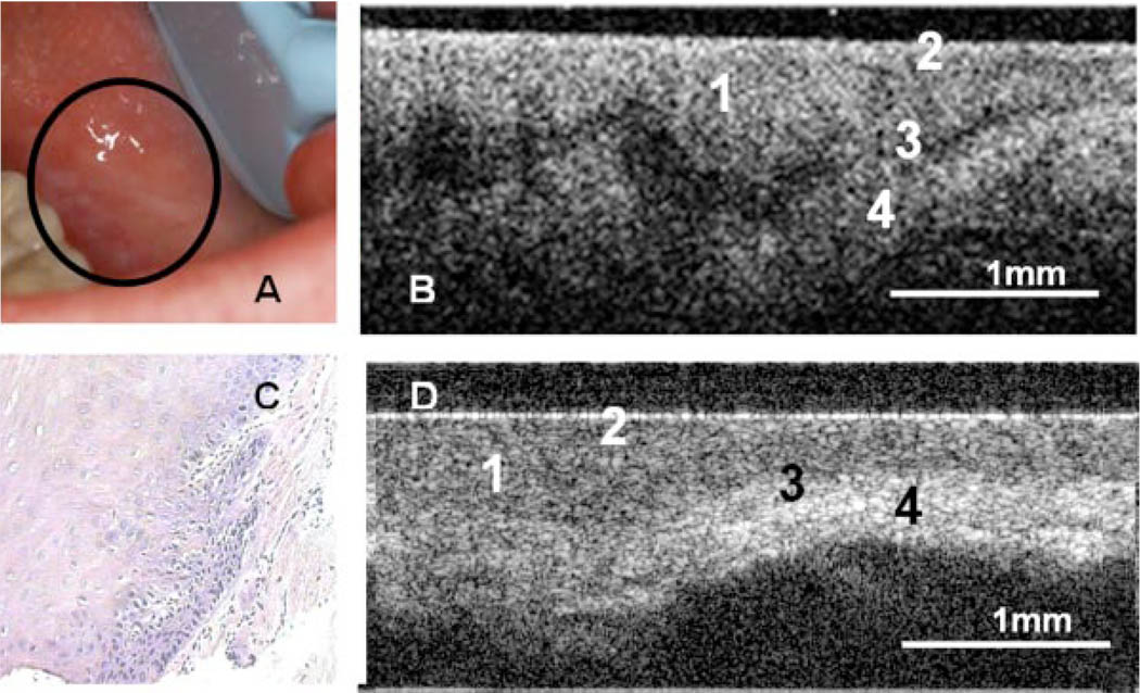
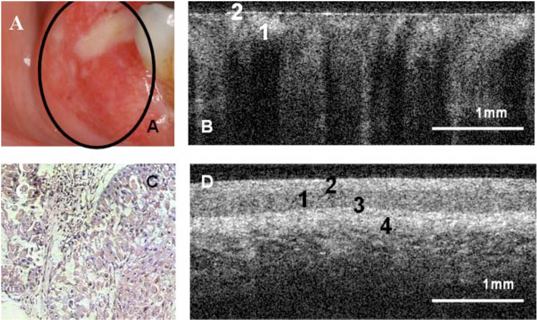
In vivo, non-invasive optical coherence tomography (OCT) permits high-resolution imaging of tissue surfaces and subsurfaces, with the potential capability for detection and mapping of epithelial pathologies.
To evaluate the clinical capability of non-invasive in vivo OCT for diagnosing oral dysplasia and malignancy.
In 50 patients with oral lesions, conventional clinical examination was followed by OCT imaging, then standard biopsy and histopathology. Two blinded, pre-standardized investigators separately diagnosed each lesion based on (1) OCT and (2) histopathology.
Intra- and inter-observer agreement between diagnoses based on histopathology and imaging data was excellent, with lambda values between 0.844 and 0.896. Sensitivity and specificity were also very good.
These data demonstrate the excellent capability of in vivo OCT for detecting and diagnosing oral premalignancy and malignancy in human subjects.
在体内,非侵入性光学相干断层扫描(OCT)可对组织表面和亚表面进行高分辨率成像,具有检测和绘制上皮病变的潜在能力。
评估非侵入性体内OCT诊断口腔发育异常和恶性肿瘤的临床能力。
对50例口腔病变患者,先进行传统临床检查,然后进行OCT成像,再进行标准活检和组织病理学检查。两名经过预标准化且不知情的研究者分别基于(1)OCT和(2)组织病理学对每个病变进行诊断。
基于组织病理学和成像数据的诊断之间,观察者内和观察者间的一致性极佳,λ值在0.844至0.896之间。敏感性和特异性也非常好。
这些数据表明体内OCT在检测和诊断人类口腔癌前病变和恶性肿瘤方面具有出色的能力。