Sun Ning, Lee Andrew, Wu Joseph C
Molecular Imaging Program at Stanford (MIPS), Department of Radiology, Stanford University School of Medicine, Stanford, CA, USA.
Nat Protoc. 2009;4(8):1192-201. doi: 10.1038/nprot.2009.100. Epub 2009 Jul 23.
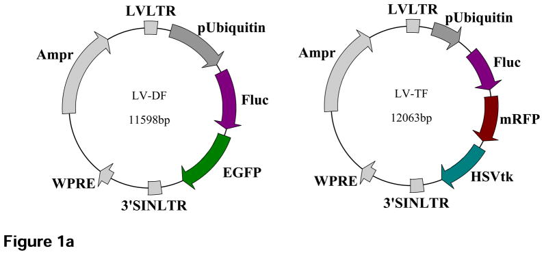
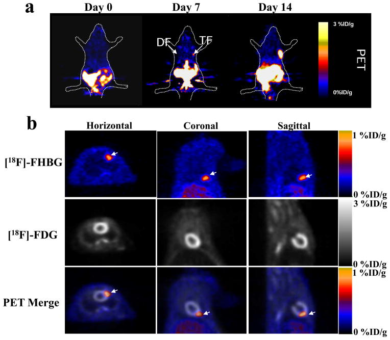
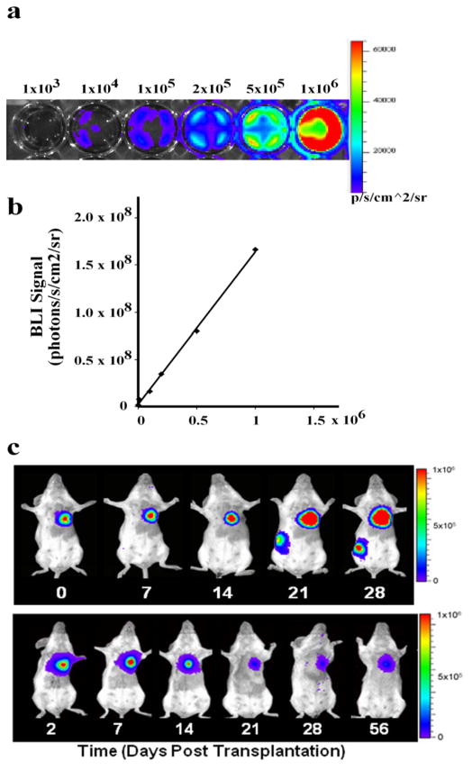
Development of non-invasive and accurate methods to track cell fate after delivery will greatly expedite transition of embryonic stem (ES) cell therapy to the clinic. In this protocol, we describe the in vivo monitoring of stem cell survival, proliferation and migration using reporter genes. We established stable ES cell lines constitutively expressing double fusion (DF; enhanced green fluorescent protein and firefly luciferase) or triple fusion (TF; monomeric red fluorescent protein, firefly luciferase and herpes simplex virus thymidine kinase (HSVtk)) reporter genes using lentiviral transduction. We used fluorescence-activated cell sorting to purify these populations in vitro, bioluminescence imaging and positron emission tomography (PET) imaging to track them in vivo and fluorescence immunostaining to confirm the results ex vivo. Unlike other methods of cell tracking, such as iron particle and radionuclide labeling, reporter genes are inherited genetically and can be used to monitor cell proliferation and survival for the lifetime of transplanted cells and their progeny.
开发无创且准确的方法来追踪细胞移植后的命运,将极大地加速胚胎干细胞(ES)治疗向临床的转化。在本方案中,我们描述了使用报告基因对干细胞存活、增殖和迁移进行体内监测的方法。我们通过慢病毒转导建立了稳定表达双融合(DF;增强型绿色荧光蛋白和萤火虫荧光素酶)或三融合(TF;单体红色荧光蛋白、萤火虫荧光素酶和单纯疱疹病毒胸苷激酶(HSVtk))报告基因的ES细胞系。我们在体外使用荧光激活细胞分选来纯化这些细胞群体,在体内使用生物发光成像和正电子发射断层扫描(PET)成像来追踪它们,并在体外使用荧光免疫染色来确认结果。与其他细胞追踪方法(如铁颗粒和放射性核素标记)不同,报告基因是通过遗传方式继承的,可用于在移植细胞及其后代的整个生命周期内监测细胞增殖和存活。