Macey Paul M, Richard Christopher A, Kumar Rajesh, Woo Mary A, Ogren Jennifer A, Avedissian Christina, Thompson Paul M, Harper Ronald M
School of Nursing, University of California Los Angeles, Los Angeles, California, United States of America.
PLoS One. 2009 Jul 30;4(7):e6436. doi: 10.1371/journal.pone.0006436.
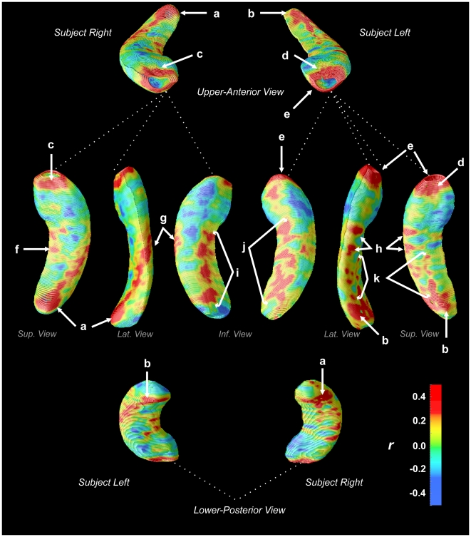
Children with congenital central hypoventilation syndrome (CCHS), a genetic disorder characterized by diminished drive to breathe during sleep and impaired CO(2) sensitivity, show brain structural and functional changes on magnetic resonance imaging (MRI) scans, with impaired responses in specific hippocampal regions, suggesting localized injury.We assessed total volume and regional variation in hippocampal surface morphology to identify areas affected in the syndrome. We studied 18 CCHS (mean age+/-std: 15.1+/-2.2 years; 8 female) and 32 healthy control (age 15.2+/-2.4 years; 14 female) children, and traced hippocampi on 1 mm(3) resolution T1-weighted scans, collected with a 3.0 Tesla MRI scanner. Regional hippocampal volume variations, adjusted for cranial volume, were compared between groups based on t-tests of surface distances to the structure midline, with correction for multiple comparisons. Significant tissue losses emerged in CCHS patients on the left side, with a trend for loss on the right; however, most areas affected on the left also showed equivalent right-sided volume reductions. Reduced regional volumes appeared in the left rostral hippocampus, bilateral areas in mid and mid-to-caudal regions, and a dorsal-caudal region, adjacent to the fimbria.The volume losses may result from hypoxic exposure following hypoventilation during sleep-disordered breathing, or from developmental or vascular consequences of genetic mutations in the syndrome. The sites of change overlap regions of abnormal functional responses to respiratory and autonomic challenges. Affected hippocampal areas have roles associated with memory, mood, and indirectly, autonomic regulation; impairments in these behavioral and physiological functions appear in CCHS.
患有先天性中枢性低通气综合征(CCHS)的儿童,这是一种遗传性疾病,其特征是睡眠期间呼吸驱动力减弱和二氧化碳敏感性受损,在磁共振成像(MRI)扫描中显示出脑结构和功能变化,特定海马区域反应受损,提示存在局部损伤。我们评估了海马表面形态的总体积和区域变化,以确定该综合征中受影响的区域。我们研究了18名CCHS儿童(平均年龄±标准差:15.1±2.2岁;8名女性)和32名健康对照儿童(年龄15.2±2.4岁;14名女性),并在使用3.0特斯拉MRI扫描仪采集的1立方毫米分辨率的T1加权扫描图像上描绘海马。基于到结构中线的表面距离的t检验,并对多重比较进行校正,比较了两组之间经颅容积校正后的海马区域体积变化。CCHS患者左侧出现明显的组织损失,右侧有损失趋势;然而,左侧受影响的大多数区域右侧体积也有同等程度的减少。左侧喙侧海马、中部和中尾区域的双侧区域以及与伞相邻的背尾区域出现区域体积减小。这些体积损失可能是由于睡眠呼吸障碍期间通气不足后的缺氧暴露,或者是该综合征中基因突变的发育或血管后果。变化部位与对呼吸和自主神经挑战的异常功能反应区域重叠。受影响的海马区域与记忆、情绪以及间接的自主神经调节有关;这些行为和生理功能的损害在CCHS中出现。