Anjomshoaa A, Nasri S, Humar B, McCall J L, Chatterjee A, Yoon H-S, McNoe L, Black M A, Reeve A E
Cancer Genetics Laboratory, Department of Biochemistry, University of Otago, Dunedin, New Zealand.
Br J Cancer. 2009 Sep 1;101(5):822-8. doi: 10.1038/sj.bjc.6605229. Epub 2009 Aug 4.
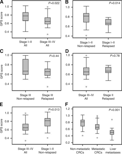
We have recently reported an inverse relationship between colon cancer progression and tumour proliferative activity. Here, we extend our findings by evaluating the proliferative activity of liver metastatic lesions and primary colorectal cancers (CRC) that differ in their metastatic potential.
Using an earlier established multi-gene proliferation signature (GPS), proliferative levels were analysed in 73 primary CRCs and 27 liver metastases.
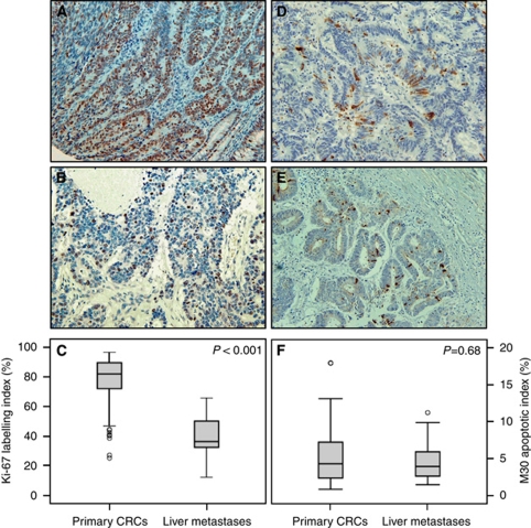
Compared with primary CRCs, we observed a significantly lower expression of the GPS in liver metastases and confirmed their lower proliferative levels by quantitative RT-PCR and Ki-67 immunostaining. No difference could be detected in apoptotic indices as assessed by M30 immunostaining, indicating that the net growth rate is lower in metastases relative to primary tumours. Notably, relapsed primaries or those with established metastases had significantly lower proliferative activity than CRCs that were non-metastatic and did not relapse.
Our results suggest that slow proliferation is a biological characteristic of both liver metastases and those primary tumours with the ability to metastasise. The delineation of the mechanisms underlying the inverse association between proliferation and CRC aggressiveness may be important for the development of new therapeutic strategies.
我们最近报道了结肠癌进展与肿瘤增殖活性之间的负相关关系。在此,我们通过评估具有不同转移潜能的肝转移瘤和原发性结直肠癌(CRC)的增殖活性来扩展我们的研究结果。
使用先前建立的多基因增殖特征(GPS),分析了73例原发性CRC和27例肝转移瘤的增殖水平。
与原发性CRC相比,我们观察到肝转移瘤中GPS的表达显著降低,并通过定量RT-PCR和Ki-67免疫染色证实其增殖水平较低。通过M30免疫染色评估的凋亡指数没有差异,表明相对于原发性肿瘤,转移瘤中的净生长率较低。值得注意的是,复发的原发性肿瘤或已发生转移的肿瘤的增殖活性明显低于未发生转移且未复发的CRC。
我们的结果表明,增殖缓慢是肝转移瘤和具有转移能力的原发性肿瘤的生物学特征。阐明增殖与CRC侵袭性之间负相关的潜在机制可能对新治疗策略的开发很重要。