Freifeld Oren, Greenspan Hayit, Goldberger Jacob
Department of Biomedical Engineering, Tel-Aviv University, Tel Aviv 69978, Israel.
Int J Biomed Imaging. 2009;2009:715124. doi: 10.1155/2009/715124. Epub 2009 Sep 10.
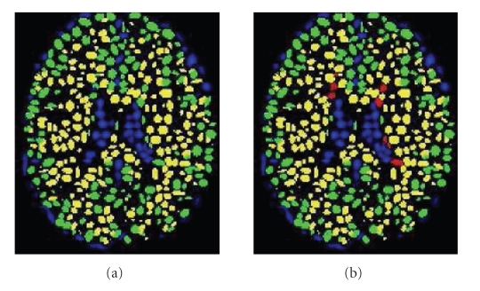
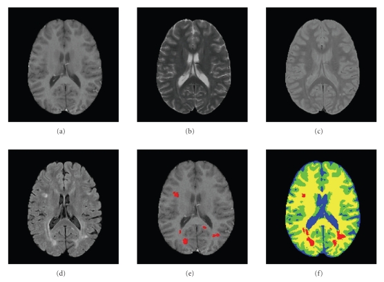
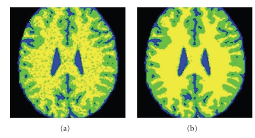
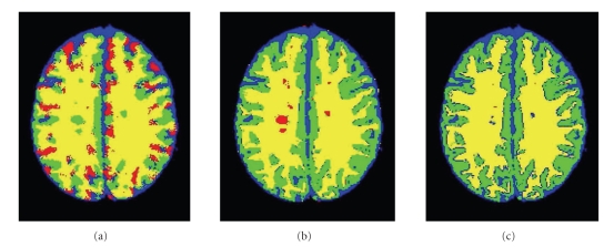
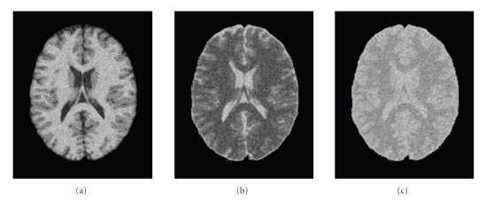
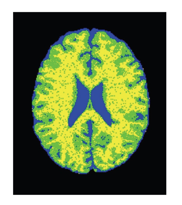
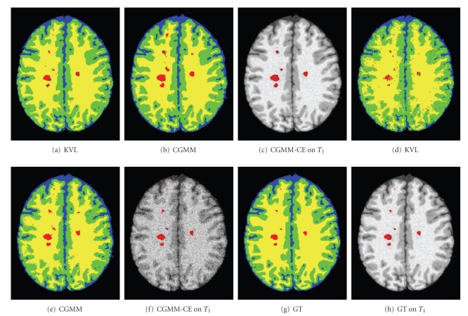
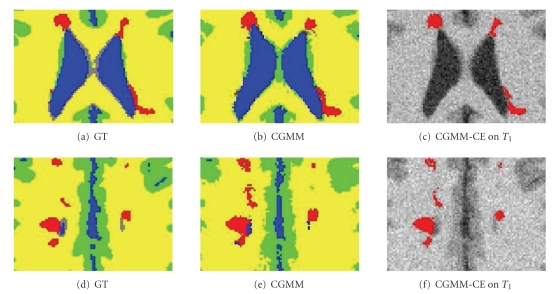
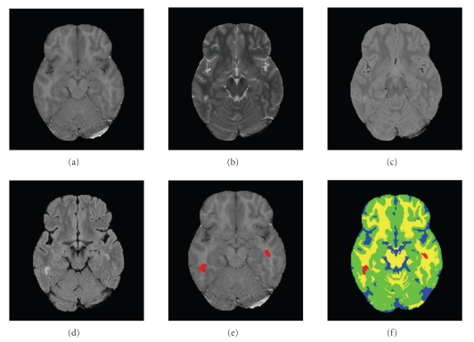
This paper focuses on the detection and segmentation of Multiple Sclerosis (MS) lesions in magnetic resonance (MRI) brain images. To capture the complex tissue spatial layout, a probabilistic model termed Constrained Gaussian Mixture Model (CGMM) is proposed based on a mixture of multiple spatially oriented Gaussians per tissue. The intensity of a tissue is considered a global parameter and is constrained, by a parameter-tying scheme, to be the same value for the entire set of Gaussians that are related to the same tissue. MS lesions are identified as outlier Gaussian components and are grouped to form a new class in addition to the healthy tissue classes. A probability-based curve evolution technique is used to refine the delineation of lesion boundaries. The proposed CGMM-CE algorithm is used to segment 3D MRI brain images with an arbitrary number of channels. The CGMM-CE algorithm is automated and does not require an atlas for initialization or parameter learning. Experimental results on both standard brain MRI simulation data and real data indicate that the proposed method outperforms previously suggested approaches, especially for highly noisy data.
本文聚焦于磁共振(MRI)脑图像中多发性硬化(MS)病灶的检测与分割。为了捕捉复杂的组织空间布局,基于每个组织多个空间定向高斯混合,提出了一种称为约束高斯混合模型(CGMM)的概率模型。组织的强度被视为一个全局参数,并通过参数绑定方案,被约束为与同一组织相关的整个高斯集合具有相同的值。MS病灶被识别为异常高斯分量,并除了健康组织类别之外,被分组形成一个新类别。基于概率的曲线演化技术用于细化病灶边界的描绘。所提出的CGMM-CE算法用于分割具有任意通道数的3D MRI脑图像。CGMM-CE算法是自动化的,不需要图谱进行初始化或参数学习。在标准脑MRI模拟数据和真实数据上的实验结果表明,所提出的方法优于先前提出的方法,特别是对于高噪声数据。