• 文献检索
  • 文档翻译
  • 深度研究
  • 学术资讯
  • Suppr Zotero 插件Zotero 插件
  • 邀请有礼
  • 套餐&价格
  • 历史记录
应用&插件
Suppr Zotero 插件Zotero 插件浏览器插件Mac 客户端Windows 客户端微信小程序
定价
高级版会员购买积分包购买API积分包
服务
文献检索文档翻译深度研究API 文档MCP 服务
关于我们
关于 Suppr公司介绍联系我们用户协议隐私条款
关注我们

Suppr 超能文献

核心技术专利:CN118964589B侵权必究
粤ICP备2023148730 号-1Suppr @ 2026

文献检索

告别复杂PubMed语法,用中文像聊天一样搜索,搜遍4000万医学文献。AI智能推荐,让科研检索更轻松。

立即免费搜索

文件翻译

保留排版,准确专业,支持PDF/Word/PPT等文件格式,支持 12+语言互译。

免费翻译文档

深度研究

AI帮你快速写综述,25分钟生成高质量综述,智能提取关键信息,辅助科研写作。

立即免费体验

基于补丁的空间一致性分割:在脑磁共振成像中多发性硬化症病变的应用

Patch-Based Segmentation with Spatial Consistency: Application to MS Lesions in Brain MRI.

作者信息

Mechrez Roey, Goldberger Jacob, Greenspan Hayit

机构信息

Biomedical Engineering Department, Tel-Aviv University, 69978 Tel Aviv, Israel.

Engineering Faculty, Bar-Ilan University, 52900 Ramat Gan, Israel.

出版信息

Int J Biomed Imaging. 2016;2016:7952541. doi: 10.1155/2016/7952541. Epub 2016 Jan 24.

DOI:10.1155/2016/7952541
PMID:26904103
原文链接:https://pmc.ncbi.nlm.nih.gov/articles/PMC4745344/
Abstract

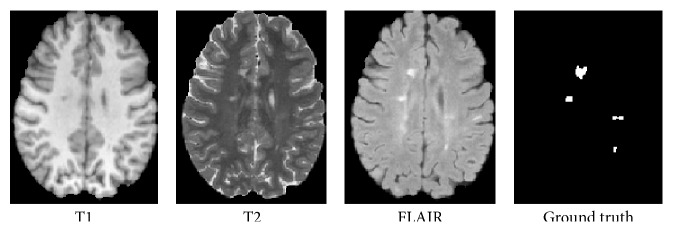
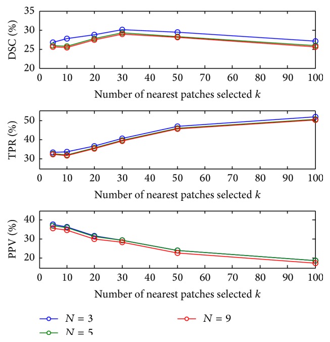
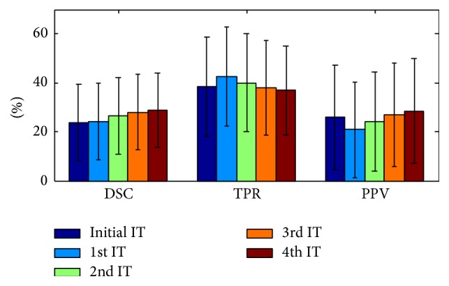
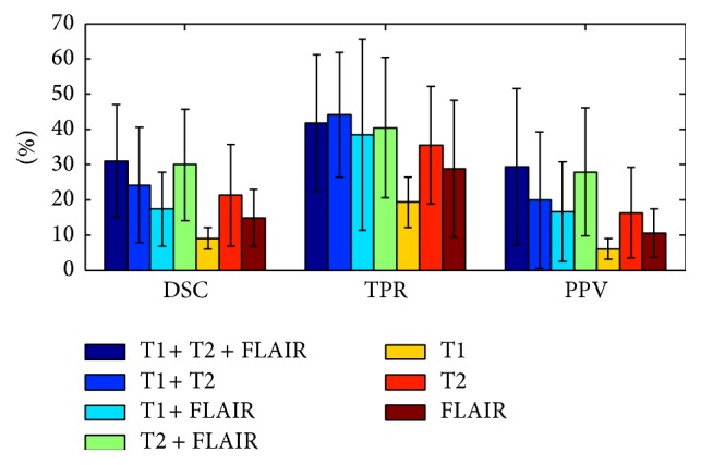
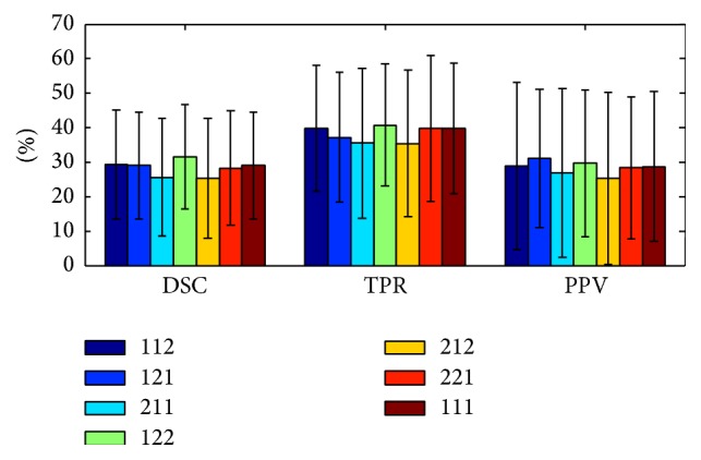
This paper presents an automatic lesion segmentation method based on similarities between multichannel patches. A patch database is built using training images for which the label maps are known. For each patch in the testing image, k similar patches are retrieved from the database. The matching labels for these k patches are then combined to produce an initial segmentation map for the test case. Finally an iterative patch-based label refinement process based on the initial segmentation map is performed to ensure the spatial consistency of the detected lesions. The method was evaluated in experiments on multiple sclerosis (MS) lesion segmentation in magnetic resonance images (MRI) of the brain. An evaluation was done for each image in the MICCAI 2008 MS lesion segmentation challenge. Results are shown to compete with the state of the art in the challenge. We conclude that the proposed algorithm for segmentation of lesions provides a promising new approach for local segmentation and global detection in medical images.

摘要

本文提出了一种基于多通道图像块相似性的自动病变分割方法。利用已知标签图的训练图像构建一个图像块数据库。对于测试图像中的每个图像块,从数据库中检索出k个相似的图像块。然后将这k个图像块的匹配标签进行合并,以生成测试病例的初始分割图。最后,基于初始分割图执行基于图像块的迭代标签细化过程,以确保检测到的病变的空间一致性。该方法在脑部磁共振成像(MRI)的多发性硬化症(MS)病变分割实验中进行了评估。对2008年医学图像计算方法与计算机辅助干预国际会议(MICCAI)MS病变分割挑战赛中的每幅图像都进行了评估。结果表明该方法在挑战赛中与当前的先进技术具有竞争力。我们得出结论,所提出的病变分割算法为医学图像中的局部分割和全局检测提供了一种有前景的新方法。

https://cdn.ncbi.nlm.nih.gov/pmc/blobs/f0a9/4745344/af3a5fd5108a/IJBI2016-7952541.alg.001.jpg
https://cdn.ncbi.nlm.nih.gov/pmc/blobs/f0a9/4745344/04b95cedfe95/IJBI2016-7952541.001.jpg
https://cdn.ncbi.nlm.nih.gov/pmc/blobs/f0a9/4745344/652de1930981/IJBI2016-7952541.002.jpg
https://cdn.ncbi.nlm.nih.gov/pmc/blobs/f0a9/4745344/087f7f9fb7ea/IJBI2016-7952541.003.jpg
https://cdn.ncbi.nlm.nih.gov/pmc/blobs/f0a9/4745344/34494a2ecb37/IJBI2016-7952541.004.jpg
https://cdn.ncbi.nlm.nih.gov/pmc/blobs/f0a9/4745344/44ded6758c7b/IJBI2016-7952541.005.jpg
https://cdn.ncbi.nlm.nih.gov/pmc/blobs/f0a9/4745344/3f3a82463df3/IJBI2016-7952541.006.jpg
https://cdn.ncbi.nlm.nih.gov/pmc/blobs/f0a9/4745344/30b905de3c1e/IJBI2016-7952541.007.jpg
https://cdn.ncbi.nlm.nih.gov/pmc/blobs/f0a9/4745344/3033e3fd2096/IJBI2016-7952541.008.jpg
https://cdn.ncbi.nlm.nih.gov/pmc/blobs/f0a9/4745344/9b2f0739ecb6/IJBI2016-7952541.009.jpg
https://cdn.ncbi.nlm.nih.gov/pmc/blobs/f0a9/4745344/f7451051f7cf/IJBI2016-7952541.010.jpg
https://cdn.ncbi.nlm.nih.gov/pmc/blobs/f0a9/4745344/af3a5fd5108a/IJBI2016-7952541.alg.001.jpg

相似文献

1
Patch-Based Segmentation with Spatial Consistency: Application to MS Lesions in Brain MRI.基于补丁的空间一致性分割:在脑磁共振成像中多发性硬化症病变的应用
Int J Biomed Imaging. 2016;2016:7952541. doi: 10.1155/2016/7952541. Epub 2016 Jan 24.
2
Neuro-fuzzy patch-wise R-CNN for multiple sclerosis segmentation.基于神经模糊补丁 R-CNN 的多发性硬化分割。
Med Biol Eng Comput. 2020 Sep;58(9):2161-2175. doi: 10.1007/s11517-020-02225-6. Epub 2020 Jul 17.
3
Hierarchical multi-atlas label fusion with multi-scale feature representation and label-specific patch partition.具有多尺度特征表示和特定标签块划分的分层多图谱标签融合
Neuroimage. 2015 Feb 1;106:34-46. doi: 10.1016/j.neuroimage.2014.11.025. Epub 2014 Nov 20.
4
Prostate cancer segmentation from MRI by a multistream fusion encoder.基于多流融合编码器的 MRI 前列腺癌分割。
Med Phys. 2023 Sep;50(9):5489-5504. doi: 10.1002/mp.16374. Epub 2023 Apr 5.
5
Automatic segmentation of white matter hyperintensities: validation and comparison with state-of-the-art methods on both Multiple Sclerosis and elderly subjects.自动分割脑白质高信号:在多发性硬化症和老年患者中验证和比较最先进的方法。
Neuroimage Clin. 2022;33:102940. doi: 10.1016/j.nicl.2022.102940. Epub 2022 Jan 10.
6
Limited One-time Sampling Irregularity Map (LOTS-IM) for Automatic Unsupervised Assessment of White Matter Hyperintensities and Multiple Sclerosis Lesions in Structural Brain Magnetic Resonance Images.用于结构磁共振成像中白质高信号和多发性硬化病变自动无监督评估的有限一次性采样不规则图(LOTS-IM)。
Comput Med Imaging Graph. 2020 Jan;79:101685. doi: 10.1016/j.compmedimag.2019.101685. Epub 2019 Nov 27.
7
Improving the detection of new lesions in multiple sclerosis with a cascaded 3D fully convolutional neural network approach.采用级联3D全卷积神经网络方法提高多发性硬化症新病灶的检测能力。
Front Neurosci. 2022 Nov 24;16:1007619. doi: 10.3389/fnins.2022.1007619. eCollection 2022.
8
Rotation-invariant multi-contrast non-local means for MS lesion segmentation.用于多发性硬化症病变分割的旋转不变多对比度非局部均值法
Neuroimage Clin. 2015 May 13;8:376-89. doi: 10.1016/j.nicl.2015.05.001. eCollection 2015.
9
New lesion segmentation for multiple sclerosis brain images with imaging and lesion-aware augmentation.基于成像和病灶感知增强技术的多发性硬化症脑部图像新病灶分割方法
Front Neurosci. 2022 Oct 21;16:1007453. doi: 10.3389/fnins.2022.1007453. eCollection 2022.
10
Automatic iterative segmentation of multiple sclerosis lesions using Student's t mixture models and probabilistic anatomical atlases in FLAIR images.利用学生t混合模型和概率性解剖图谱在液体衰减反转恢复(FLAIR)图像中对多发性硬化病变进行自动迭代分割
Comput Biol Med. 2016 Jun 1;73:10-23. doi: 10.1016/j.compbiomed.2016.03.025. Epub 2016 Apr 1.

引用本文的文献

1
A Hybrid Intelligence Approach for Circulating Tumor Cell Enumeration in Digital Pathology by Using CNN and Weak Annotations.一种利用卷积神经网络(CNN)和弱标注进行数字病理学中循环肿瘤细胞计数的混合智能方法。
IEEE Access. 2023;11:142992-143003. doi: 10.1109/access.2023.3343701. Epub 2023 Dec 18.
2
Inpainting surgical occlusion from laparoscopic video sequences for robot-assisted interventions.从腹腔镜视频序列中修复手术遮挡以用于机器人辅助干预
J Med Imaging (Bellingham). 2023 Jul;10(4):045002. doi: 10.1117/1.JMI.10.4.045002. Epub 2023 Aug 29.
3
Automatic brain lesion segmentation on standard magnetic resonance images: a scoping review.

本文引用的文献

1
Multiple sclerosis lesion segmentation using dictionary learning and sparse coding.基于字典学习和稀疏编码的多发性硬化症病灶分割
Med Image Comput Comput Assist Interv. 2013;16(Pt 1):735-42. doi: 10.1007/978-3-642-40811-3_92.
2
Integration of sparse multi-modality representation and anatomical constraint for isointense infant brain MR image segmentation.用于等强度婴儿脑磁共振图像分割的稀疏多模态表示与解剖学约束的整合
Neuroimage. 2014 Apr 1;89:152-64. doi: 10.1016/j.neuroimage.2013.11.040. Epub 2013 Nov 28.
3
Temporally consistent probabilistic detection of new multiple sclerosis lesions in brain MRI.
基于标准磁共振图像的脑自动病变分割:范围综述。
BMJ Open. 2021 Jan 29;11(1):e042660. doi: 10.1136/bmjopen-2020-042660.
4
Pathomechanisms of HIV-Associated Cerebral Small Vessel Disease: A Comprehensive Clinical and Neuroimaging Protocol and Analysis Pipeline.HIV 相关脑小血管疾病的发病机制:一项全面的临床和神经影像学方案及分析流程
Front Neurol. 2020 Dec 15;11:595463. doi: 10.3389/fneur.2020.595463. eCollection 2020.
5
U-NetPlus: A Modified Encoder-Decoder U-Net Architecture for Semantic and Instance Segmentation of Surgical Instruments from Laparoscopic Images.U-NetPlus:一种用于从腹腔镜图像中对手术器械进行语义和实例分割的改进型编码器-解码器U-Net架构。
Annu Int Conf IEEE Eng Med Biol Soc. 2019 Jul;2019:7205-7211. doi: 10.1109/EMBC.2019.8856791.
6
Automated Segmentation of Tissues Using CT and MRI: A Systematic Review.基于 CT 和 MRI 的组织自动分割:系统评价。
Acad Radiol. 2019 Dec;26(12):1695-1706. doi: 10.1016/j.acra.2019.07.006. Epub 2019 Aug 10.
7
Multiple Sclerosis Lesion Segmentation Using Joint Label Fusion.使用联合标签融合的多发性硬化病变分割
Patch Based Tech Med Imaging (2017). 2017 Sep;10530:138-145. doi: 10.1007/978-3-319-67434-6_16. Epub 2017 Aug 31.
8
Baby brain atlases.婴儿脑图谱。
Neuroimage. 2019 Jan 15;185:865-880. doi: 10.1016/j.neuroimage.2018.04.003. Epub 2018 Apr 3.
9
Longitudinal multiple sclerosis lesion segmentation: Resource and challenge.纵向多发性硬化病变分割:资源与挑战。
Neuroimage. 2017 Mar 1;148:77-102. doi: 10.1016/j.neuroimage.2016.12.064. Epub 2017 Jan 11.
10
A Semiautomatic Method for Multiple Sclerosis Lesion Segmentation on Dual-Echo MR Imaging: Application in a Multicenter Context.一种用于双回波磁共振成像的多发性硬化病变分割的半自动方法:在多中心环境中的应用。
AJNR Am J Neuroradiol. 2016 Nov;37(11):2043-2049. doi: 10.3174/ajnr.A4874. Epub 2016 Jul 21.
脑 MRI 中新多发性硬化病变的时间一致概率检测。
IEEE Trans Med Imaging. 2013 Aug;32(8):1490-503. doi: 10.1109/TMI.2013.2258403. Epub 2013 Apr 16.
4
Efficient brain lesion segmentation using multi-modality tissue-based feature selection and support vector machines.基于多模态组织特征选择和支持向量机的高效脑损伤分割。
Int J Numer Method Biomed Eng. 2013 Sep;29(9):905-15. doi: 10.1002/cnm.2537. Epub 2013 Jan 10.
5
Review of automatic segmentation methods of multiple sclerosis white matter lesions on conventional magnetic resonance imaging.磁共振常规成像多发性硬化脑白质病变自动分割方法的研究进展。
Med Image Anal. 2013 Jan;17(1):1-18. doi: 10.1016/j.media.2012.09.004. Epub 2012 Sep 29.
6
3D Slicer as an image computing platform for the Quantitative Imaging Network.3D Slicer 作为定量成像网络的图像计算平台。
Magn Reson Imaging. 2012 Nov;30(9):1323-41. doi: 10.1016/j.mri.2012.05.001. Epub 2012 Jul 6.
7
Stochastic relaxation, gibbs distributions, and the bayesian restoration of images.随机松弛,吉布斯分布,以及贝叶斯图像恢复。
IEEE Trans Pattern Anal Mach Intell. 1984 Jun;6(6):721-41. doi: 10.1109/tpami.1984.4767596.
8
Automatic detection of gadolinium-enhancing multiple sclerosis lesions in brain MRI using conditional random fields.基于条件随机场的脑 MRI 钆增强多发性硬化病变的自动检测
IEEE Trans Med Imaging. 2012 Jun;31(6):1181-94. doi: 10.1109/TMI.2012.2186639. Epub 2012 Feb 3.
9
An automated tool for detection of FLAIR-hyperintense white-matter lesions in Multiple Sclerosis.用于检测多发性硬化症中 FLAIR 高信号白质病变的自动化工具。
Neuroimage. 2012 Feb 15;59(4):3774-83. doi: 10.1016/j.neuroimage.2011.11.032. Epub 2011 Nov 18.
10
A Cellular Neural Network methodology for the automated segmentation of multiple sclerosis lesions.一种用于自动分割多发性硬化病变的细胞神经网络方法。
J Neurosci Methods. 2012 Jan 15;203(1):193-9. doi: 10.1016/j.jneumeth.2011.08.047. Epub 2011 Sep 7.