College of Life Sciences, Zhejiang University, Key Laboratory for Cell and Gene Engineering of Zhejiang Province, Hangzhou, PR China.
J Cell Mol Med. 2010 Jun;14(6B):1494-508. doi: 10.1111/j.1582-4934.2009.00912.x. Epub 2009 Sep 24.
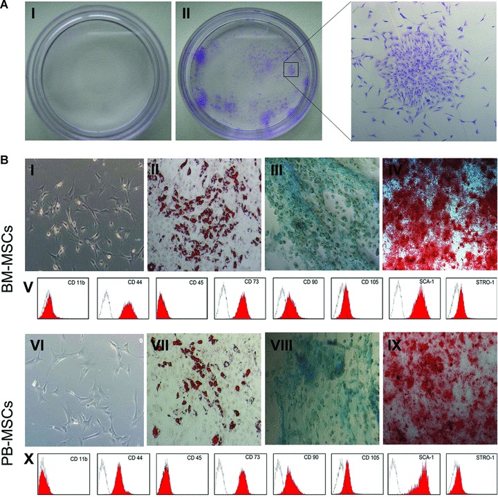
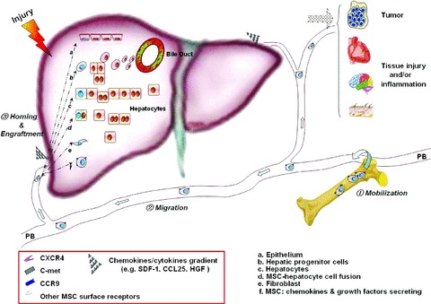
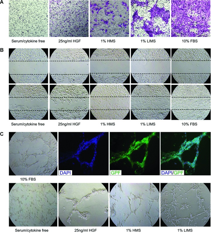
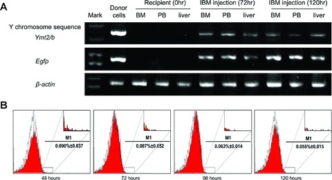
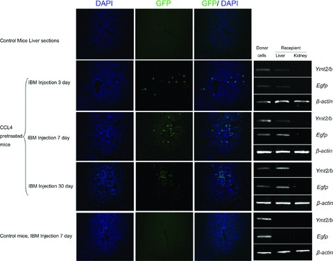
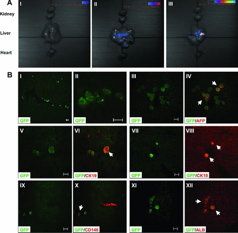
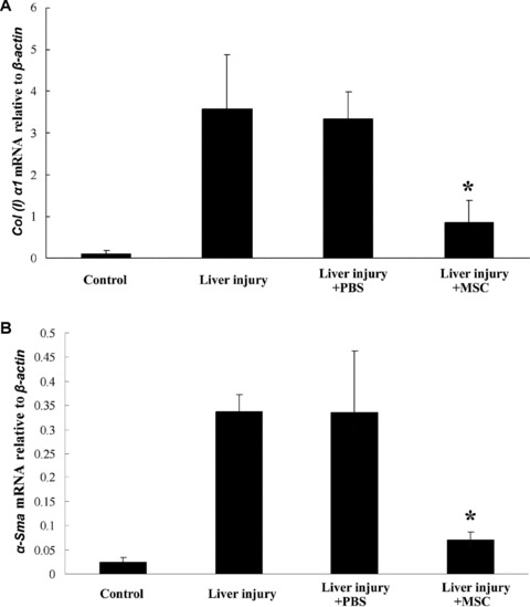
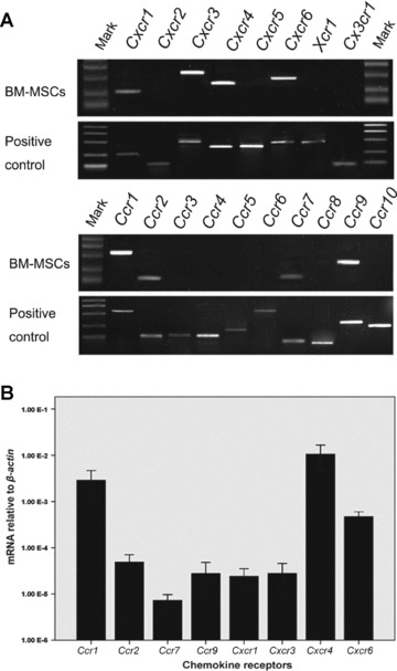
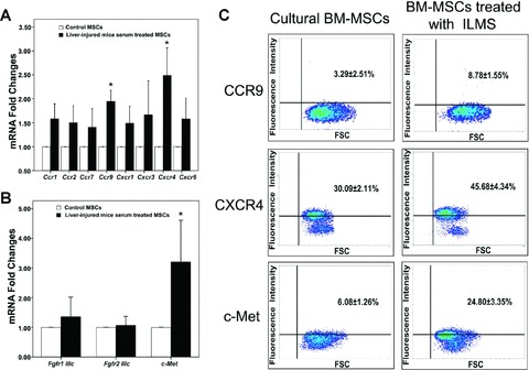
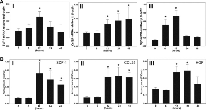
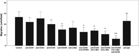
Recent studies suggest that mesenchymal stem cells (MSCs) possess a greater differentiation potential than once thought and that they have the capacity to regenerate damaged tissues/organs. However, the evidence is insufficient, and the mechanism governing the recruitment and homing of MSCs to these injured sites is not well understood. We first examined the MSCs circulating in peripheral blood and then performed chemotaxis, wound healing and tubule-formation assays to investigate the migration capability of mouse bone marrow MSCs (mBM-MSCs) in response to liver-injury signals. In addition, BM-MSCs from donor enhanced green fluorescent protein transgenic male mice were transplanted into liver-injured co-isogenic female recipients, either by intra-bone marrow injection or through the caudal vein, to allow in vivo tracking analysis of the cell fate after transplantation. Donor-derived cells were analysed by in vivo imaging analysis, PCR, flow cytometry and frozen sections. Microarray and real-time PCR were used for chemokine/cytokine and receptor analyses. We successfully isolated circulating MSCs in peripheral blood of liver-injured mice and provided direct evidence that mBM-MSCs could be mobilized into the circulation and recruited into the liver after stimulation of liver injury. CCR9, CXCR4 and c-MET were essential for directing cellular migration towards the injured liver. The recruited mBM-MSCs may play different roles, including hepatic fate specification and down-regulation of the activity of hepatic stellate cells which inhibits over-accumulation of collagen and development of liver fibrosis. Our results provide new insights into liver repair involving endogenous BM-MSCs and add new information for consideration when developing clinical protocols involving the MSCs.
最近的研究表明,间充质干细胞(MSCs)比人们之前认为的具有更大的分化潜力,并且它们具有再生受损组织/器官的能力。然而,证据还不充分,调节 MSC 募集和归巢到这些受损部位的机制还不是很清楚。我们首先检查了外周血中的 MSC,然后进行趋化、伤口愈合和管形成测定,以研究对肝损伤信号响应的小鼠骨髓 MSC(mBM-MSCs)的迁移能力。此外,来自供体增强型绿色荧光蛋白转基因雄性小鼠的 BM-MSCs 被移植到肝损伤同基因雌性受体内,通过骨髓内注射或通过尾静脉,允许在体内对移植后细胞命运进行跟踪分析。通过体内成像分析、PCR、流式细胞术和冷冻切片分析供体细胞。微阵列和实时 PCR 用于趋化因子/细胞因子和受体分析。我们成功地从肝损伤小鼠的外周血中分离出循环 MSC,并提供了直接证据,表明 mBM-MSCs 可以在肝损伤刺激后动员到循环中并募集到肝脏中。CCR9、CXCR4 和 c-MET 对于指导细胞向受损肝脏的迁移是必不可少的。募集的 mBM-MSCs 可能发挥不同的作用,包括肝命运特化和下调肝星状细胞的活性,抑制胶原过度积累和肝纤维化的发展。我们的研究结果为涉及内源性 BM-MSCs 的肝修复提供了新的见解,并为开发涉及 MSC 的临床方案提供了新的信息。