• 文献检索
  • 文档翻译
  • 深度研究
  • 学术资讯
  • Suppr Zotero 插件Zotero 插件
  • 邀请有礼
  • 套餐&价格
  • 历史记录
应用&插件
Suppr Zotero 插件Zotero 插件浏览器插件Mac 客户端Windows 客户端微信小程序
定价
高级版会员购买积分包购买API积分包
服务
文献检索文档翻译深度研究API 文档MCP 服务
关于我们
关于 Suppr公司介绍联系我们用户协议隐私条款
关注我们

Suppr 超能文献

核心技术专利:CN118964589B侵权必究
粤ICP备2023148730 号-1Suppr @ 2026

文献检索

告别复杂PubMed语法,用中文像聊天一样搜索,搜遍4000万医学文献。AI智能推荐,让科研检索更轻松。

立即免费搜索

文件翻译

保留排版,准确专业,支持PDF/Word/PPT等文件格式,支持 12+语言互译。

免费翻译文档

深度研究

AI帮你快速写综述,25分钟生成高质量综述,智能提取关键信息,辅助科研写作。

立即免费体验

用于小动物成像和性能评估的锥形束微计算机断层扫描系统。

Cone beam micro-CT system for small animal imaging and performance evaluation.

作者信息

Zhu Shouping, Tian Jie, Yan Guorui, Qin Chenghu, Feng Jinchao

机构信息

Medical Image Processing Group, Institute of Automation, Chinese Academy of Sciences, Beijing 100190, China.

出版信息

Int J Biomed Imaging. 2009;2009:960573. doi: 10.1155/2009/960573. Epub 2009 Sep 22.

DOI:10.1155/2009/960573
PMID:19794829
原文链接:https://pmc.ncbi.nlm.nih.gov/articles/PMC2754077/
Abstract

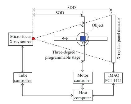
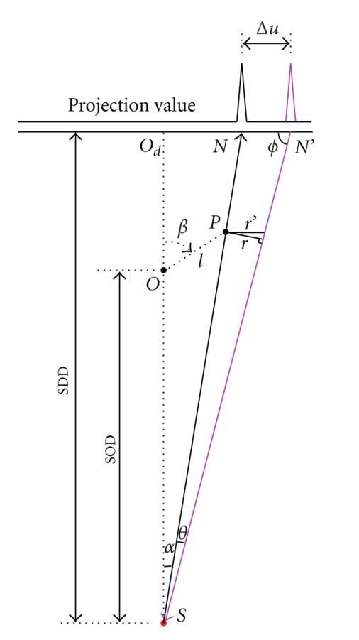
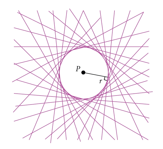
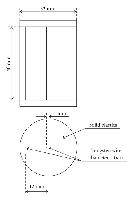
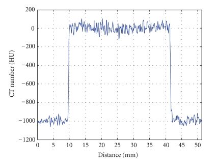
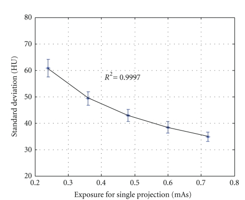
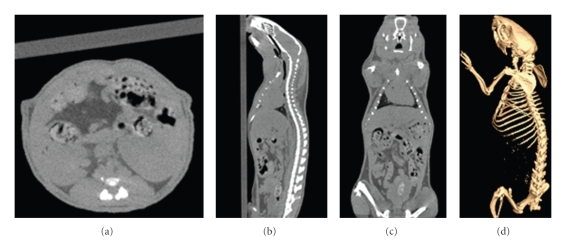
A prototype cone-beam micro-CT system for small animal imaging has been developed by our group recently, which consists of a microfocus X-ray source, a three-dimensional programmable stage with object holder, and a flat-panel X-ray detector. It has a large field of view (FOV), which can acquire the whole body imaging of a normal-size mouse in a single scan which usually takes about several minutes or tens of minutes. FDK method is adopted for 3D reconstruction with Graphics Processing Unit (GPU) acceleration. In order to reconstruct images with high spatial resolution and low artifacts, raw data preprocessing and geometry calibration are implemented before reconstruction. A method which utilizes a wire phantom to estimate the residual horizontal offset of the detector is proposed, and 1D point spread function is used to assess the performance of geometric calibration quantitatively. System spatial resolution, image uniformity and noise, and low contrast resolution have been studied. Mouse images with and without contrast agent are illuminated in this paper. Experimental results show that the system is suitable for small animal imaging and is adequate to provide high-resolution anatomic information for bioluminescence tomography to build a dual modality system.

摘要

我们团队最近开发了一种用于小动物成像的锥形束微CT系统原型,它由一个微焦点X射线源、一个带有样品架的三维可编程平台和一个平板X射线探测器组成。它具有大视野(FOV),可以在一次扫描中获取正常大小小鼠的全身成像,通常需要几分钟或几十分钟。采用FDK方法进行三维重建,并通过图形处理单元(GPU)加速。为了重建具有高空间分辨率和低伪影的图像,在重建前进行原始数据预处理和几何校准。提出了一种利用线模体估计探测器残余水平偏移的方法,并使用一维点扩散函数定量评估几何校准的性能。研究了系统的空间分辨率、图像均匀性和噪声以及低对比度分辨率。本文展示了有和没有造影剂的小鼠图像。实验结果表明,该系统适用于小动物成像,足以提供高分辨率的解剖信息用于生物发光断层成像以构建双模态系统。

https://cdn.ncbi.nlm.nih.gov/pmc/blobs/0898/2754077/cbc7823f2081/IJBI2009-960573.0012.jpg
https://cdn.ncbi.nlm.nih.gov/pmc/blobs/0898/2754077/a3088d8ec5aa/IJBI2009-960573.001.jpg
https://cdn.ncbi.nlm.nih.gov/pmc/blobs/0898/2754077/77fcb5b0a423/IJBI2009-960573.002.jpg
https://cdn.ncbi.nlm.nih.gov/pmc/blobs/0898/2754077/fdf930375d96/IJBI2009-960573.003.jpg
https://cdn.ncbi.nlm.nih.gov/pmc/blobs/0898/2754077/1a1d4f089f71/IJBI2009-960573.004.jpg
https://cdn.ncbi.nlm.nih.gov/pmc/blobs/0898/2754077/fb9842c601f8/IJBI2009-960573.005.jpg
https://cdn.ncbi.nlm.nih.gov/pmc/blobs/0898/2754077/67fd8f6af0c8/IJBI2009-960573.006.jpg
https://cdn.ncbi.nlm.nih.gov/pmc/blobs/0898/2754077/d951365c7edb/IJBI2009-960573.007.jpg
https://cdn.ncbi.nlm.nih.gov/pmc/blobs/0898/2754077/b7fa3b678296/IJBI2009-960573.008.jpg
https://cdn.ncbi.nlm.nih.gov/pmc/blobs/0898/2754077/5a3e6bb0eb0a/IJBI2009-960573.009.jpg
https://cdn.ncbi.nlm.nih.gov/pmc/blobs/0898/2754077/73ec2d2fe8d6/IJBI2009-960573.0010.jpg
https://cdn.ncbi.nlm.nih.gov/pmc/blobs/0898/2754077/89c033cd911a/IJBI2009-960573.0011.jpg
https://cdn.ncbi.nlm.nih.gov/pmc/blobs/0898/2754077/cbc7823f2081/IJBI2009-960573.0012.jpg

相似文献

1
Cone beam micro-CT system for small animal imaging and performance evaluation.用于小动物成像和性能评估的锥形束微计算机断层扫描系统。
Int J Biomed Imaging. 2009;2009:960573. doi: 10.1155/2009/960573. Epub 2009 Sep 22.
2
Accurate technique for complete geometric calibration of cone-beam computed tomography systems.用于锥束计算机断层扫描系统完全几何校准的精确技术。
Med Phys. 2005 Apr;32(4):968-83. doi: 10.1118/1.1869652.
3
ALBIRA: a small animal PET∕SPECT∕CT imaging system.ALBIRA:小动物 PET/SPECT/CT 成像系统。
Med Phys. 2013 May;40(5):051906. doi: 10.1118/1.4800798.
4
Flat-panel cone-beam computed tomography for image-guided radiation therapy.用于图像引导放射治疗的平板锥形束计算机断层扫描
Int J Radiat Oncol Biol Phys. 2002 Aug 1;53(5):1337-49. doi: 10.1016/s0360-3016(02)02884-5.
5
A prototype table-top inverse-geometry volumetric CT system.一种桌面式反几何容积CT系统原型。
Med Phys. 2006 Jun;33(6):1867-78. doi: 10.1118/1.2192887.
6
Fast and effective single-scan dual-energy cone-beam CT reconstruction and decomposition denoising based on dual-energy vectorization.基于双能矢量化的快速有效单扫描双能锥形束 CT 重建和分解去噪。
Med Phys. 2021 Sep;48(9):4843-4856. doi: 10.1002/mp.15117. Epub 2021 Aug 11.
7
Stationary computed tomography with source and detector in linear symmetric geometry: Direct filtered backprojection reconstruction.直线对称几何源和探测器的静止计算机断层扫描:直接滤波反投影重建。
Med Phys. 2020 Jun;47(5):2222-2236. doi: 10.1002/mp.14058. Epub 2020 Apr 3.
8
Flat panel detector-based cone beam computed tomography with a circle-plus-two-arcs data acquisition orbit: preliminary phantom study.基于平板探测器的采用圆加双弧数据采集轨道的锥束计算机断层扫描:初步体模研究
Med Phys. 2003 Jul;30(7):1694-705. doi: 10.1118/1.1582470.
9
Cone-beam imaging with tilted rotation axis: Method and performance evaluation.倾斜旋转轴的锥束成像:方法与性能评估。
Med Phys. 2020 Aug;47(8):3305-3320. doi: 10.1002/mp.14209. Epub 2020 May 22.
10
Towards the clinical implementation of iterative low-dose cone-beam CT reconstruction in image-guided radiation therapy: cone/ring artifact correction and multiple GPU implementation.迈向迭代低剂量锥形束CT重建在图像引导放射治疗中的临床应用:锥形/环形伪影校正与多GPU实现
Med Phys. 2014 Nov;41(11):111912. doi: 10.1118/1.4898324.

引用本文的文献

1
Dual and Multi-Target Cone-Beam X-ray Luminescence Computed Tomography Based on the DeepCB-XLCT Network.基于深度锥束X射线发光计算机断层扫描网络的双目标和多目标锥束X射线发光计算机断层扫描
Bioengineering (Basel). 2024 Aug 28;11(9):874. doi: 10.3390/bioengineering11090874.
2
X-ray scintillator lens-coupled with CMOS camera for pre-clinical cardiac vascular imaging-A feasibility study.X 射线闪烁体透镜与 CMOS 相机耦合在临床前心血管成像中的应用——一项可行性研究。
PLoS One. 2022 Feb 11;17(2):e0262913. doi: 10.1371/journal.pone.0262913. eCollection 2022.
3
Comparison of GPU reconstruction based on different symmetries for dual-head PET.

本文引用的文献

1
In vivo mouse studies with bioluminescence tomography.利用生物发光断层扫描技术进行的小鼠体内研究。
Opt Express. 2006 Aug 21;14(17):7801-9. doi: 10.1364/oe.14.007801.
2
A novel software platform for medical image processing and analyzing.一种用于医学图像处理和分析的新型软件平台。
IEEE Trans Inf Technol Biomed. 2008 Nov;12(6):800-12. doi: 10.1109/TITB.2008.926395.
3
Multimodality molecular imaging.多模态分子成像
双探头 PET 基于不同对称性的 GPU 重建比较。
Med Phys. 2019 Jun;46(6):2696-2708. doi: 10.1002/mp.13529. Epub 2019 May 8.
4
Regularized reconstruction based on joint L and total variation for sparse-view cone-beam X-ray luminescence computed tomography.基于联合L范数和全变差的正则化重建用于稀疏视图锥束X射线发光计算机断层扫描
Biomed Opt Express. 2018 Dec 3;10(1):1-17. doi: 10.1364/BOE.10.000001. eCollection 2019 Jan 1.
5
Self-assembled micro-computed tomography for dental education.自组装微计算机断层扫描在口腔医学教育中的应用。
PLoS One. 2018 Dec 26;13(12):e0209698. doi: 10.1371/journal.pone.0209698. eCollection 2018.
6
Sparse Reconstruction of Fluorescence Molecular Tomography Using Variable Splitting and Alternating Direction Scheme.基于变量分裂和交替方向法的荧光分子断层成像稀疏重建。
Mol Imaging Biol. 2018 Feb;20(1):37-46. doi: 10.1007/s11307-017-1088-4.
7
Fast and Robust Reconstruction for Fluorescence Molecular Tomography via Regularization.基于正则化的荧光分子断层成像快速稳健重建
Biomed Res Int. 2016;2016:5065217. doi: 10.1155/2016/5065217. Epub 2016 Dec 6.
8
Cone Beam X-ray Luminescence Computed Tomography Based on Bayesian Method.基于贝叶斯方法的锥形束X射线发光计算机断层扫描
IEEE Trans Med Imaging. 2017 Jan;36(1):225-235. doi: 10.1109/TMI.2016.2603843. Epub 2016 Aug 26.
9
Novel l 2,1-norm optimization method for fluorescence molecular tomography reconstruction.用于荧光分子断层成像重建的新型l 2,1 -范数优化方法
Biomed Opt Express. 2016 May 23;7(6):2342-59. doi: 10.1364/BOE.7.002342. eCollection 2016 Jun 1.
10
L Regularization for Bioluminescence Tomography Based on the Split Bregman Method.基于分裂Bregman方法的生物发光断层成像的L正则化
Mol Imaging Biol. 2016 Dec;18(6):830-837. doi: 10.1007/s11307-016-0970-9.
IEEE Eng Med Biol Mag. 2008 Sep-Oct;27(5):48-57. doi: 10.1109/MEMB.2008.923962.
4
In vivo small-animal imaging using micro-CT and digital subtraction angiography.使用微型计算机断层扫描(micro-CT)和数字减影血管造影术进行体内小动物成像。
Phys Med Biol. 2008 Oct 7;53(19):R319-50. doi: 10.1088/0031-9155/53/19/R01. Epub 2008 Aug 29.
5
An optimization-based method for geometrical calibration in cone-beam CT without dedicated phantoms.一种用于锥束CT中无需专用体模的基于优化的几何校准方法。
Phys Med Biol. 2008 Jul 21;53(14):3841-61. doi: 10.1088/0031-9155/53/14/009. Epub 2008 Jun 26.
6
Imaging in the era of molecular oncology.分子肿瘤学时代的影像学
Nature. 2008 Apr 3;452(7187):580-9. doi: 10.1038/nature06917.
7
Image reconstruction from cone-beam projections: necessary and sufficient conditions and reconstruction methods.从锥束投影重建图像:必要和充分条件及重建方法。
IEEE Trans Med Imaging. 1985;4(1):14-25. doi: 10.1109/TMI.1985.4307689.
8
A quality assurance phantom for the performance evaluation of volumetric micro-CT systems.用于容积式微型计算机断层扫描系统性能评估的质量保证体模。
Phys Med Biol. 2007 Dec 7;52(23):7087-108. doi: 10.1088/0031-9155/52/23/021. Epub 2007 Nov 15.
9
A microPET/CT system for in vivo small animal imaging.一种用于活体小动物成像的微型正电子发射断层显像/计算机断层扫描(microPET/CT)系统。
Phys Med Biol. 2007 Jul 7;52(13):3881-94. doi: 10.1088/0031-9155/52/13/015. Epub 2007 Jun 4.
10
A geometric calibration method for cone beam CT systems.一种用于锥束CT系统的几何校准方法。
Med Phys. 2006 Jun;33(6):1695-706. doi: 10.1118/1.2198187.