Terrovitis John, Lautamäki Riikka, Bonios Michael, Fox James, Engles James M, Yu Jianhua, Leppo Michelle K, Pomper Martin G, Wahl Richard L, Seidel Jurgen, Tsui Benjamin M, Bengel Frank M, Abraham M Roselle, Marbán Eduardo
The Heart Institute, Cedars Sinai Medical Center, Los Angeles, California 90048, USA.
J Am Coll Cardiol. 2009 Oct 20;54(17):1619-26. doi: 10.1016/j.jacc.2009.04.097.
The aim of this study was to quantify acute myocardial retention of cardiac-derived stem cells (CDCs) and evaluate different delivery methods with positron emission tomography (PET).
Success of stem cell transplantation for cardiac regeneration is partially limited by low retention/engraftment of the delivered cells. A clinically applicable method for accurate quantification of cell retention would enable optimization of cell delivery.
The CDCs were derived from syngeneic, male Wistar Kyoto (WK) rats labeled with [(18)F]-fluoro-deoxy-glucose ((18)FDG) and injected intramyocardially into the ischemic region of female WK rats after permanent left coronary artery ligation. The effects of fibrin glue (FG), bradycardia (adenosine), and cardiac arrest were examined. Imaging with (18)FDG PET was performed for quantification of cell retention. Quantitative polymerase chain reaction (PCR) for the male-specific SRY gene was performed to validate the PET results.
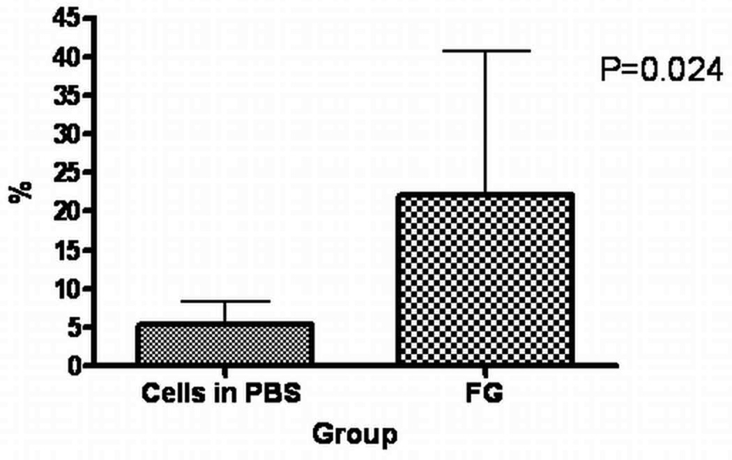
Myocardial retention of cells suspended in phosphate-buffered saline 1 h after delivery was 17.6 +/- 11.5% by PCR and 17.8 +/- 7.3% by PET. When CDCs were injected immediately after induction of cardiac arrest, retention was increased to 75.6 +/- 18.6%. Adenosine slowed the ventricular rate and doubled CDC retention (35.4 +/- 5.3%). A similar increase in CDC retention was observed after epicardial application of FG at the injection site (37.5 +/- 8.2%). The PCR revealed a significant increase in 3-week cell engraftment in the FG animals (22.1 +/- 18.6% and 5.3 +/- 3.1%, for FG and phosphate-buffered saline, respectively).
In vivo PET permits accurate measurement of CDC retention early after intramyocardial delivery. Sealing injection sites with FG or lowering ventricular rate by adenosine might be clinically translatable methods for improving stem cell engraftment in a beating heart.
本研究旨在通过正电子发射断层扫描(PET)对心脏来源干细胞(CDC)的急性心肌滞留情况进行定量分析,并评估不同的递送方法。
心脏再生的干细胞移植成功率部分受限于所递送细胞的低滞留/植入率。一种临床适用的准确量化细胞滞留的方法将有助于优化细胞递送。
CDC取自同基因雄性Wistar Kyoto(WK)大鼠,用[(18)F]-氟脱氧葡萄糖((18)FDG)标记,在永久性左冠状动脉结扎后经心肌内注射到雌性WK大鼠的缺血区域。研究了纤维蛋白胶(FG)、心动过缓(腺苷)和心脏骤停的影响。采用(18)FDG PET成像对细胞滞留情况进行定量分析。对雄性特异性SRY基因进行定量聚合酶链反应(PCR)以验证PET结果。
递送后1小时,悬浮于磷酸盐缓冲盐水中的细胞经PCR检测心肌滞留率为17.6±11.5%,经PET检测为17.8±7.3%。在心脏骤停诱导后立即注射CDC,滞留率增加到75.6±18.6%。腺苷减慢心室率,使CDC滞留率加倍(35.4±5.3%)。在注射部位的心外膜应用FG后,观察到CDC滞留率有类似增加(37.5±8.2%)。PCR显示FG组动物在3周时细胞植入有显著增加(FG组和磷酸盐缓冲盐水组分别为22.1±18.6%和5.3±3.1%)。
体内PET可在心肌内递送后早期准确测量CDC滞留情况。用FG封闭注射部位或用腺苷降低心室率可能是临床上可转化的改善跳动心脏中干细胞植入的方法。