Elkahwaji J E, Hauke R J, Brawner C M
Department of Internal Medicine, Section of Adult Oncology and Hematology and GU Oncology Research Laboratory, University of Nebraska Medical Center, Omaha, NE 68198-7680, USA.
Br J Cancer. 2009 Nov 17;101(10):1740-8. doi: 10.1038/sj.bjc.6605370. Epub 2009 Oct 20.
Although the aetiology of prostate cancer remains unknown, we hypothesised that chronic bacterial insult has a major role in prostate carcinogenesis.
Male C3H/HeOuJ mice, infected with phosphate-buffered saline or Escherichia coli bacteria, were killed at 5 days, or at 12 or 26 weeks. Harvested prostate tissues were evaluated for inflammatory responses and immunostained for neoplastic transformation markers.
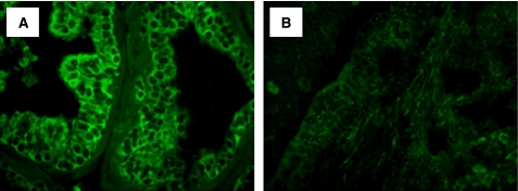
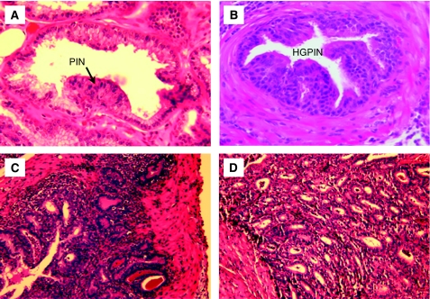
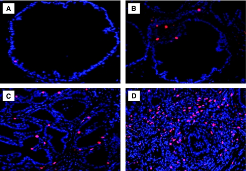
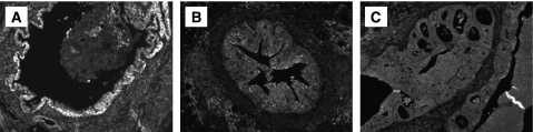
All infected mice developed bacterial prostatitis. Control mice had no prostate infections or inflammation. Mice infected for 5 days showed foci of acute inflammation with infiltrating neutrophils and epithelial necrotic debris in the prostatic glandular lumen. All mice infected for 12 weeks had evidence of chronic inflammation with dense inflammatory infiltrates in the stroma. The prostatic epithelium showed varying degrees of atypical hyperplasia with increased epithelial cell layers and cytological atypia. At 26 weeks, the dysplastic changes were more pronounced and mimicked a prostatic intraepithelial neoplasia and high-grade dysplasia. Prostatic glands exhibiting reactive dysplasia had a stronger staining for oxidative DNA damage, increased epithelial cell proliferation, and a decrease in androgen receptor, GSTP1, p27(Kip1), and PTEN expression, when compared with control prostate glands.
These data demonstrate that chronic inflammation induces focal prostatic glandular atypia and suggest a potential linkage between inflammation and prostatic neoplasia.
尽管前列腺癌的病因尚不清楚,但我们推测慢性细菌感染在前列腺癌发生过程中起主要作用。
将感染磷酸盐缓冲盐水或大肠杆菌的雄性C3H/HeOuJ小鼠在5天、12周或26周时处死。对收获的前列腺组织进行炎症反应评估,并对肿瘤转化标志物进行免疫染色。
所有感染小鼠均发生细菌性前列腺炎。对照小鼠无前列腺感染或炎症。感染5天的小鼠前列腺腺腔内可见急性炎症灶,有浸润的中性粒细胞和上皮坏死碎片。所有感染12周的小鼠均有慢性炎症证据,间质中有密集的炎症浸润。前列腺上皮显示不同程度的非典型增生,上皮细胞层数增加且有细胞学非典型性。在26周时,发育异常变化更为明显,类似前列腺上皮内瘤变和高级别发育异常。与对照前列腺相比,表现为反应性发育异常的前列腺腺体对氧化性DNA损伤染色更强,上皮细胞增殖增加,雄激素受体、GSTP1、p27(Kip1)和PTEN表达减少。
这些数据表明慢性炎症可诱导前列腺腺泡局灶性异型增生,并提示炎症与前列腺肿瘤之间存在潜在联系。