Jansen Jacobus F A, Schöder Heiko, Lee Nancy Y, Wang Ya, Pfister David G, Fury Matthew G, Stambuk Hilda E, Humm John L, Koutcher Jason A, Shukla-Dave Amita
Department of Medical Physics, Memorial Sloan-Kettering Cancer Center, New York, NY 10065, USA.
Int J Radiat Oncol Biol Phys. 2010 Aug 1;77(5):1403-10. doi: 10.1016/j.ijrobp.2009.07.009. Epub 2009 Nov 10.
To assess noninvasively the tumor microenvironment of neck nodal metastases in patients with head-and-neck cancer by investigating the relationship between tumor perfusion measured using dynamic contrast-enhanced magnetic resonance imaging (DCE-MRI) and hypoxia measured by (18)F-fluoromisonidazole ((18)F-FMISO) positron emission tomography (PET).
Thirteen newly diagnosed head-and-neck cancer patients with metastatic neck nodes underwent DCE-MRI and (18)F-FMISO PET imaging before chemotherapy and radiotherapy. The matched regions of interests from both modalities were analyzed. To examine the correlations between DCE-MRI parameters and standard uptake value (SUV) measurements from (18)F-FMISO PET, the nonparametric Spearman correlation coefficient was calculated. Furthermore, DCE-MRI parameters were compared between nodes with (18)F-FMISO uptake and nodes with no (18)F-FMISO uptake using Mann-Whitney U tests.
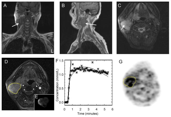
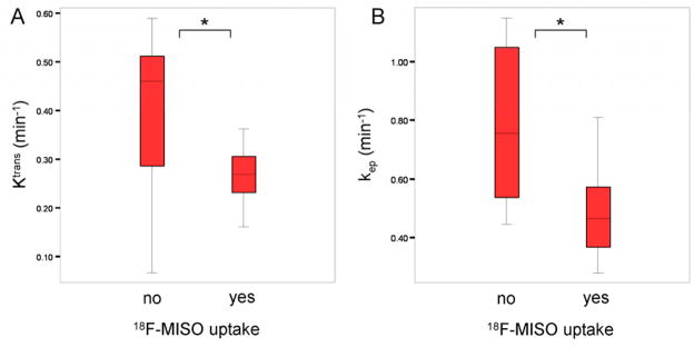
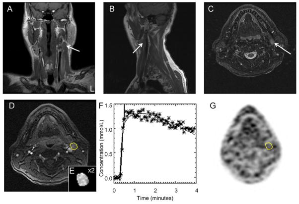
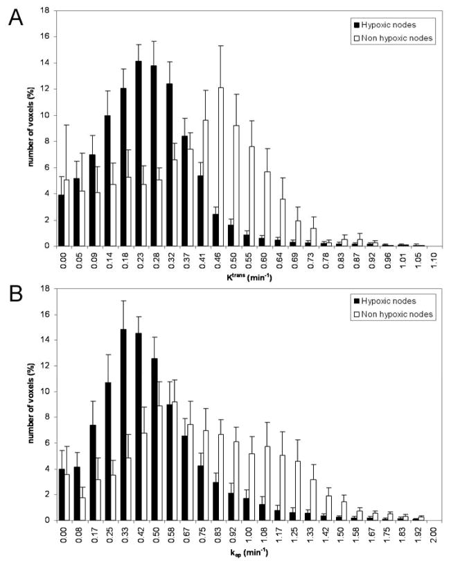
For the 13 patients, a total of 18 nodes were analyzed. The nodal size strongly correlated with the (18)F-FMISO SUV (rho = 0.74, p < 0.001). There was a strong negative correlation between the median k(ep) (redistribution rate constant) value (rho = -0.58, p = 0.042) and the (18)F-FMISO SUV. Hypoxic nodes (moderate to severe (18)F-FMISO uptake) had significantly lower median K(trans) (volume transfer constant) (p = 0.049) and median k(ep) (p = 0.027) values than did nonhypoxic nodes (no (18)F-FMISO uptake).
This initial evaluation of the preliminary results support the hypothesis that in metastatic neck lymph nodes, hypoxic nodes are poorly perfused (i.e., have significantly lower K(trans) and k(ep) values) compared with nonhypoxic nodes.
通过研究使用动态对比增强磁共振成像(DCE-MRI)测量的肿瘤灌注与用(18)F-氟米索硝唑((18)F-FMISO)正电子发射断层扫描(PET)测量的缺氧之间的关系,对头颈癌患者颈部淋巴结转移的肿瘤微环境进行无创评估。
13例新诊断的伴有颈部淋巴结转移的头颈癌患者在化疗和放疗前接受了DCE-MRI和(18)F-FMISO PET成像。对两种检查方式匹配的感兴趣区域进行分析。为了检验DCE-MRI参数与(18)F-FMISO PET的标准摄取值(SUV)测量值之间的相关性,计算了非参数Spearman相关系数。此外,使用Mann-Whitney U检验比较了有(18)F-FMISO摄取的淋巴结和无(18)F-FMISO摄取的淋巴结之间的DCE-MRI参数。
对13例患者共分析了18个淋巴结。淋巴结大小与(18)F-FMISO SUV密切相关(rho = 0.74,p < 0.001)。中位k(ep)(再分布速率常数)值与(18)F-FMISO SUV之间存在强负相关(rho = -0.58,p = 0.042)。缺氧淋巴结(中度至重度(18)F-FMISO摄取)的中位K(trans)(容积转运常数)(p = 0.049)和中位k(ep)(p = 0.027)值显著低于非缺氧淋巴结(无(18)F-FMISO摄取)。
对初步结果的这一初步评估支持以下假设:在转移性颈部淋巴结中,与非缺氧淋巴结相比,缺氧淋巴结灌注不良(即K(trans)和k(ep)值显著更低)。