Kawaguchi Amu, Nakaya Hiroyuki, Okabe Takahiro, Tensho Keiji, Nawata Masashi, Eguchi Yoshitaka, Imai Yuuki, Takaoka Kunio, Wakitani Shigeyuki
Department of Orthopaedic Surgery, Shinshu University School of Medicine, Matsumoto, Japan.
Acta Orthop. 2009 Oct;80(5):606-11. doi: 10.3109/17453670903350115.
Osteochondral defects have a limited capacity for repair. We therefore investigated the effects of tumor necrosis factor (TNF) signal blockade by etanercept (human recombinant soluble TNF receptor) on the repair of osteochondral defects in rabbit knees.
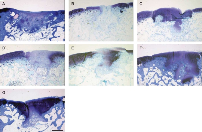
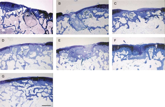
Osteochondral defects (5 mm in diameter) were created in the femoral patellar groove in rabbits. Soon after the procedure, a first subcutaneous injection of etanercept was performed. This single injection or, alternatively, 4 injections in total (twice a week for 2 weeks) were given. Each of these 2 groups was divided further into 3 subgroups: a low-dose group (0.05 microg/kg), an intermediate-dose group (0.4 microg/kg), and a high-dose group (1.6 microg /kg) with 19 rabbits in each. As a control, 19 rabbits were injected with water alone. The rabbits in each subgroup were killed 4 weeks (6 rabbits), 8 weeks (6 rabbits), or 24 weeks (7 rabbits) after surgery and repair was assessed histologically.
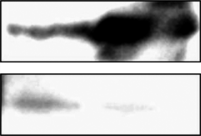
Histological examination revealed that the natural process of repair of the osteochondral defects was promoted by 4 subcutaneous injections of intermediate-dose etanercept and by 1 or 4 injections of high-dose etanercept at the various time points examined postoperatively (4, 8, and 24 weeks). Western blot showed that rabbit TNFalpha had a high affinity for etanercept.
Blocking of TNF by etanercept enabled repair of osteochondral defects in rabbit knee. Anti-TNF therapy could be a strategy for the use of tissue engineering for bone and cartilage repair.
骨软骨缺损的修复能力有限。因此,我们研究了依那西普(人重组可溶性肿瘤坏死因子受体)阻断肿瘤坏死因子(TNF)信号对兔膝关节骨软骨缺损修复的影响。
在兔股骨髌沟制造骨软骨缺损(直径5毫米)。术后不久,首次皮下注射依那西普。给予单次注射或总共4次注射(每周两次,共2周)。这两组中的每组又进一步分为3个亚组:低剂量组(0.05微克/千克)、中剂量组(0.4微克/千克)和高剂量组(1.6微克/千克),每组19只兔。作为对照,19只兔仅注射水。每个亚组的兔在术后4周(6只兔)、8周(6只兔)或24周(7只兔)处死,并进行组织学评估修复情况。
组织学检查显示,在术后检查的各个时间点(4周、8周和24周),皮下注射4次中剂量依那西普以及注射1次或4次高剂量依那西普可促进骨软骨缺损的自然修复过程。蛋白质印迹法显示兔TNFα与依那西普具有高亲和力。
依那西普阻断TNF可使兔膝关节骨软骨缺损得到修复。抗TNF治疗可能是一种用于骨和软骨修复的组织工程策略。