Wershil B K, Wang Z S, Gordon J R, Galli S J
Department of Pathology, Beth Israel Hospital, Boston, Massachusetts 02215.
J Clin Invest. 1991 Feb;87(2):446-53. doi: 10.1172/JCI115016.
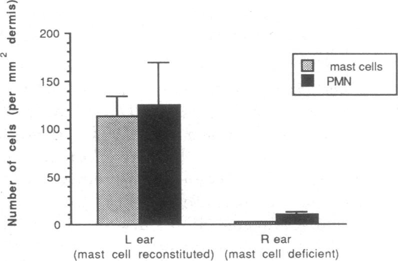
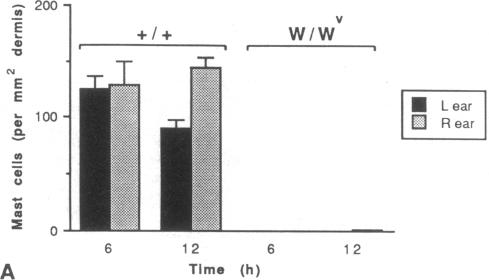
Much of the clinically important pathology associated with IgE-dependent disorders is thought to reflect the actions of the blood-borne leukocytes recruited during these responses. To evaluate the extent to which mast cells are responsible for the leukocyte infiltration associated with IgE-dependent cutaneous reactions, we attempted to elicit these responses in normal mice, genetically mast cell-deficient W/Wv mice, and in W/Wv mice selectively repaired of their mast cell deficiency by the intradermal injection of cultured mast cells derived from the congenic normal (+/+) mice. We found that the tissue swelling associated with IgE-dependent passive cutaneous anaphylaxis reactions developed rapidly and diminished markedly from 2 to 4 h after antigen challenge, but remained detectable for at least 24 h after elicitation of the responses. Infiltration of leukocytes (predominantly neutrophils) also occurred at these sites, but reached maximal levels 6-12 h after antigen challenge, persisted at high levels for 24 h, and largely waned by 48 h. Virtually all of the tissue swelling and leukocyte infiltration associated with IgE-dependent cutaneous reactions was mast cell dependent. Intradermal injection of 40 U of recombinant murine TNF-alpha (rmTNF-alpha) elicited neutrophil infiltration similar in magnitude and kinetics to that observed after IgE-dependent mast cell degranulation. A rabbit anti-rmTNF-alpha (R anti-rmTNF-alpha) antiserum, which was able to inhibit 84% of the neutrophil infiltration observed after i.d. injection of rmTNF-alpha, inhibited IgE-, and mast cell-dependent leukocyte infiltration by 47 +/- 7% in three separate experiments. These findings indicate that TNF-alpha contributes to mast cell-dependent recruitment of leukocytes during IgE-dependent cutaneous late phase reactions, but suggest that other mast cell-associated mediators probably also contribute to this response.
许多与IgE依赖性疾病相关的具有临床重要性的病理学表现被认为反映了在这些反应过程中募集的血源性白细胞的作用。为了评估肥大细胞在多大程度上导致了与IgE依赖性皮肤反应相关的白细胞浸润,我们试图在正常小鼠、遗传性肥大细胞缺陷的W/Wv小鼠以及通过皮内注射来自同基因正常(+/+)小鼠的培养肥大细胞而选择性修复肥大细胞缺陷的W/Wv小鼠中引发这些反应。我们发现,与IgE依赖性被动皮肤过敏反应相关的组织肿胀迅速出现,并在抗原激发后2至4小时明显减轻,但在反应引发后至少24小时仍可检测到。白细胞(主要是中性粒细胞)也在这些部位浸润,但在抗原激发后6至12小时达到最高水平,在高水平持续24小时,并在48小时时基本消退。几乎所有与IgE依赖性皮肤反应相关的组织肿胀和白细胞浸润都依赖于肥大细胞。皮内注射40 U重组小鼠肿瘤坏死因子-α(rmTNF-α)引发的中性粒细胞浸润在程度和动力学上与IgE依赖性肥大细胞脱颗粒后观察到的相似。一种兔抗rmTNF-α(R抗-rmTNF-α)抗血清能够抑制皮内注射rmTNF-α后观察到的84%的中性粒细胞浸润,在三个独立实验中,该抗血清抑制IgE和肥大细胞依赖性白细胞浸润的程度为47±7%。这些发现表明,肿瘤坏死因子-α在IgE依赖性皮肤晚期反应中有助于肥大细胞依赖性白细胞的募集,但也表明其他与肥大细胞相关的介质可能也参与了这一反应。