Department of Pharmacology and Clinical Pharmacology, School of Medical Sciences, Faculty of Medical and Health Sciences, The University of Auckland, Auckland, New Zealand.
Mol Pain. 2009 Nov 18;5:66. doi: 10.1186/1744-8069-5-66.
Oxaliplatin and related chemotherapeutic drugs cause painful chronic peripheral neuropathies in cancer patients. We investigated changes in neuronal size profiles and neurofilament immunoreactivity in L5 dorsal root ganglion (DRG) tissue of adult female Wistar rats after multiple-dose treatment with oxaliplatin, cisplatin, carboplatin or paclitaxel.
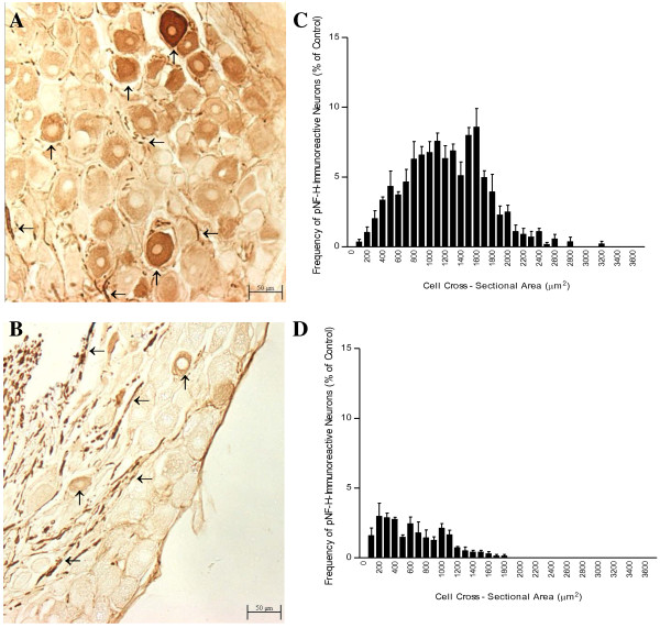
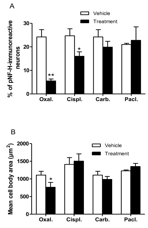
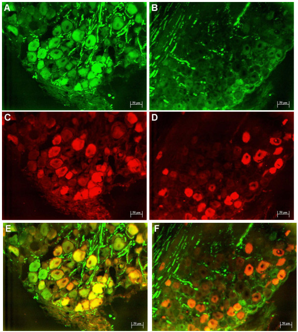
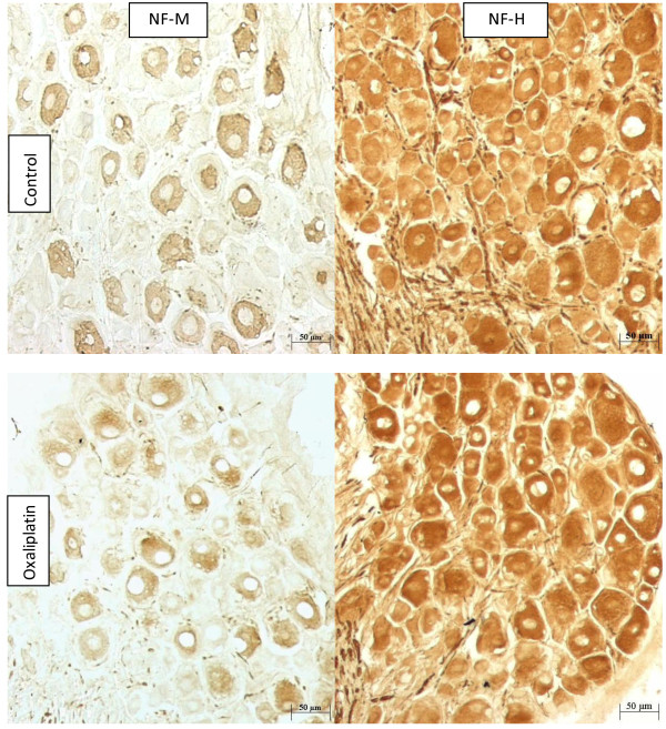
After treatment with oxaliplatin, phosphorylated neurofilament heavy subunit (pNF-H) immunoreactivity was reduced in neuronal cell bodies, but unchanged in nerve fibres, of the L5 DRG. Morphometric analysis confirmed significant changes in the number (-75%; P < 0.0002) and size (-45%; P < 0.0001) of pNF-H-immunoreactive neurons after oxaliplatin treatment. pNF-H-immunoreactive neurons had overlapping size profiles and co-localisation with neurons displaying cell body immunoreactivity for parvalbumin, non-phospho-specific neurofilament medium subunit (NF-M) and non-phospho-specific neurofilament heavy subunit (NF-H), in control DRG. However, there were no significant changes in the numbers of neurons with immunoreactivity for parvalbumin (4.6%, P = 0.82), NF-M (-1%, P = 0.96) or NF-H (0%; P = 0.93) after oxaliplatin treatment, although the sizes of parvalbumin (-29%, P = 0.047), NF-M (-11%, P = 0.038) and NF-H (-28%; P = 0.0033) immunoreactive neurons were reduced. In an independent comparison of different chemotherapeutic agents, the number of pNF-H-immunoreactive neurons was significantly altered by oxaliplatin (-77.2%; P < 0.0001) and cisplatin (-35.2%; P = 0.03) but not by carboplatin or paclitaxel, and their mean cell body area was significantly changed by oxaliplatin (-31.1%; P = 0.008) but not by cisplatin, carboplatin or paclitaxel.
This study has demonstrated a specific pattern of loss of pNF-H immunoreactivity in rat DRG tissue that corresponds with the relative neurotoxicity of oxaliplatin, cisplatin and carboplatin. Loss of pNF-H may be mechanistically linked to oxaliplatin-induced neuronal atrophy, and serves as a readily measureable endpoint of its neurotoxicity in the rat model.
奥沙利铂和相关的化疗药物会导致癌症患者出现疼痛性慢性周围神经病变。我们研究了成年雌性 Wistar 大鼠经奥沙利铂、顺铂、卡铂或紫杉醇多次治疗后 L5 背根神经节(DRG)组织中神经元大小谱和神经丝免疫反应的变化。
奥沙利铂治疗后,L5 DRG 神经元胞体中的磷酸化神经丝重链(pNF-H)免疫反应减少,但神经纤维不变。形态计量分析证实,奥沙利铂治疗后 pNF-H 免疫反应性神经元的数量减少了 75%(P < 0.0002),大小减少了 45%(P < 0.0001)。pNF-H 免疫反应性神经元的大小谱重叠,并与显示胞体免疫反应性的神经元共存,这些神经元对钙调蛋白、非磷酸化特异性神经丝中间亚基(NF-M)和非磷酸化特异性神经丝重链(NF-H)具有免疫反应性,在对照 DRG 中。然而,奥沙利铂治疗后,钙调蛋白(4.6%,P = 0.82)、NF-M(-1%,P = 0.96)或 NF-H(0%,P = 0.93)免疫反应性神经元的数量没有显著变化,尽管钙调蛋白(-29%,P = 0.047)、NF-M(-11%,P = 0.038)和 NF-H(-28%,P = 0.0033)免疫反应性神经元的大小减少。在对不同化疗药物的独立比较中,奥沙利铂(-77.2%,P < 0.0001)和顺铂(-35.2%,P = 0.03)显著改变了 pNF-H 免疫反应性神经元的数量,但卡铂或紫杉醇则没有,其平均胞体面积也因奥沙利铂而显著改变(-31.1%,P = 0.008),但不是因为顺铂、卡铂或紫杉醇。
本研究在大鼠 DRG 组织中证实了一种特定的磷酸化神经丝免疫反应缺失模式,这种缺失模式与奥沙利铂、顺铂和卡铂的相对神经毒性相对应。pNF-H 的缺失可能与奥沙利铂诱导的神经元萎缩有关,并且可以作为其在大鼠模型中神经毒性的一个易于测量的终点。