Williams Ariel A, Higgins John P T, Zhao Hongjuan, Ljunberg Börje, Brooks James D
Department of Urology, Stanford University, California, USA.
BMC Clin Pathol. 2009 Nov 18;9:9. doi: 10.1186/1472-6890-9-9.
Clear cell renal cell carcinoma (ccRCC) and chromophobe renal cell carcinoma (chRCC) can usually be distinguished by histologic characteristics. Occasionally, diagnosis proves challenging and diagnostic difficulty will likely increase as needle biopsies of renal lesions become more common.
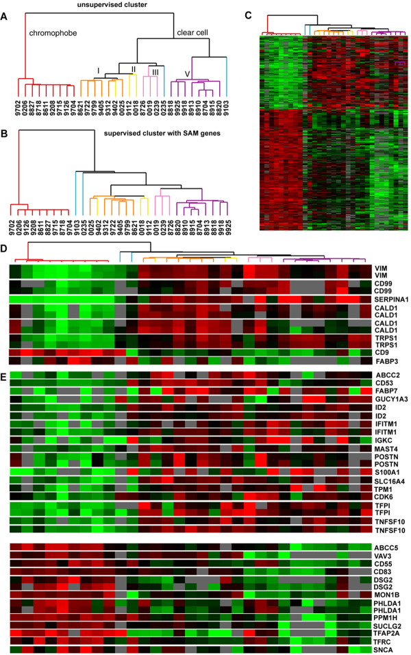
To identify markers that aid in differentiating ccRCC from chRCC, we used gene expression profiles to identify candidate markers that correlate with histology. 39 antisera and antibodies, including 35 for transcripts identified from gene expression profiling, were evaluated. Promising markers were tested on a tissue microarray (TMA) containing 428 renal neoplasms. Strength of staining of each core on the TMA was formally scored and the distribution of staining across different types of renal neoplasms was analyzed.
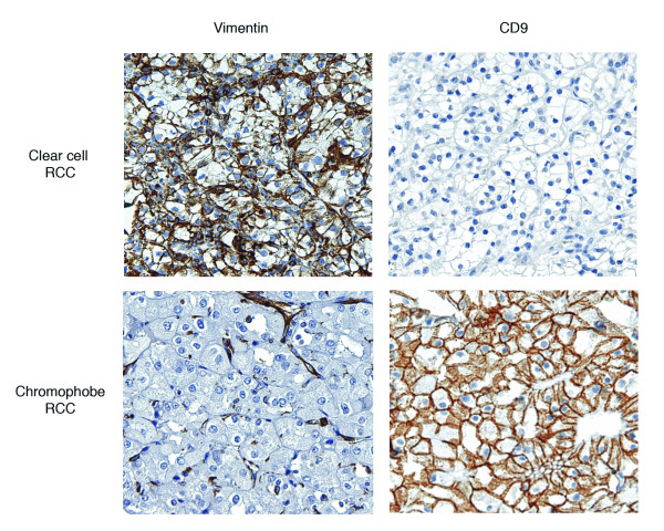
Based on results from initial immunohistochemical staining of multitissue titer arrays, 23 of the antisera and antibodies were selected for staining of the TMA. For 7 of these markers, strength of staining of each core on the TMA was formally scored. Vimentin (positive in ccRCC) and CD9 (positive in chRCC) best distinguished ccRCC from chRCC. The combination of vimentin negativity and CD9 positivity was found to distinguish chRCC from ccRCC with a sensitivity of 100.0% and a specificity of 95.2%.
Based on gene expression analysis, we identify CD9 and vimentin as candidate markers for distinguishing between ccRCC and chRCC. In difficult cases and particularly when the amount of diagnostic tissue is limited, vimentin and CD9 staining could serve as a useful adjunct in the differential diagnosis of ccRCC and chRCC.
透明细胞肾细胞癌(ccRCC)和嫌色细胞肾细胞癌(chRCC)通常可通过组织学特征加以区分。偶尔,诊断颇具挑战性,而且随着肾病变针吸活检日益普遍,诊断难度可能会增加。
为了确定有助于区分ccRCC和chRCC的标志物,我们利用基因表达谱来识别与组织学相关的候选标志物。对39种抗血清和抗体进行了评估,其中包括针对从基因表达谱中鉴定出的转录本的35种。在包含428个肾肿瘤的组织微阵列(TMA)上对有前景的标志物进行了检测。对TMA上每个核心的染色强度进行了正式评分,并分析了不同类型肾肿瘤的染色分布情况。
基于多组织滴度阵列初始免疫组化染色的结果,选择了23种抗血清和抗体对TMA进行染色。对其中7种标志物,对TMA上每个核心的染色强度进行了正式评分。波形蛋白(在ccRCC中呈阳性)和CD9(在chRCC中呈阳性)最能区分ccRCC和chRCC。发现波形蛋白阴性和CD9阳性的组合区分chRCC和ccRCC的敏感性为100.0%,特异性为95.2%。
基于基因表达分析,我们确定CD9和波形蛋白为区分ccRCC和chRCC的候选标志物。在疑难病例中,尤其是当诊断组织量有限时,波形蛋白和CD9染色可作为ccRCC和chRCC鉴别诊断的有用辅助手段。