Sadauskas Evaldas, Jacobsen Nicklas Raun, Danscher Gorm, Stoltenberg Meredin, Vogel Ulla, Larsen Agnete, Kreyling Wolfgang, Wallin Håkan
Department of Neurobiology, Institute of Anatomy, University of Aarhus, Aarhus, Denmark.
Chem Cent J. 2009 Nov 20;3:16. doi: 10.1186/1752-153X-3-16.
The fate of gold nanoparticles, 2, 40 and 100 nm, administered intratracheally to adult female mice was examined. The nanoparticles were traced by autometallography (AMG) at both ultrastructural and light microscopic levels. Also, the gold content was quantified by inductively coupled plasma mass spectrometry (ICP-MS) and neutron activation analysis (NAA). The liver is the major site of deposition of circulating gold nanoparticles. Therefore the degree of translocation was determined by the hepatic deposition of gold. Mice were instilled with 5 intratracheal doses of gold nanoparticles distributed over a period of 3 weeks and were killed 24 h after the last dose. One group of mice were given a single intratracheal dose and were killed after 1 h.
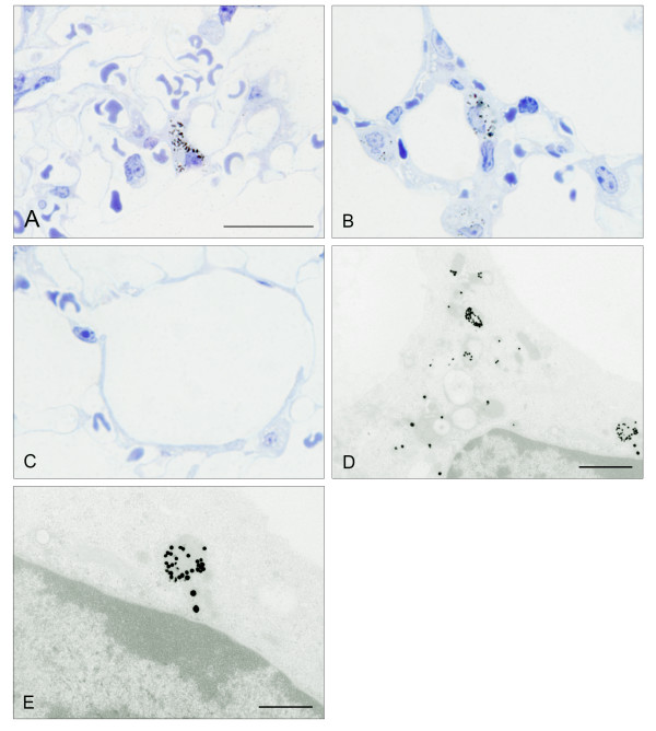
The instilled nanoparticles were found in lung macrophages already 1 h after a single instillation. In mice instilled treated repeatedly during 3 weeks, the load was substantial. Ultrastructurally, AMG silver enhanced gold nanoparticles were found in lysosome-/endosome-like organelles of the macrophages and analysis with AMG, ICP-MS and NAA of the liver revealed an almost total lack of translocation of nanoparticles. In mice given repeated instillations of 2 nm gold nanoparticles, 1.4 per thousand (by ICP-MS) to 1.9 per thousand (by NAA) of the instilled gold was detected in the liver. With the 40 nm gold, no gold was detected in the liver (detection level 2 ng, 0.1 per thousand) except for one mouse in which 3 per thousand of the instilled gold was found in the liver. No gold was detected in any liver of mice instilled with 100 nm gold (detection level 2 ng, 0.1 per thousand) except in a single animal with 0.39 per thousand of the dose in the liver.
We found that that: (1) inert gold nanoparticles, administered intratracheally are phagocytosed by lung macrophages; (2) only a tiny fraction of the gold particles is translocated into systemic circulation. (3) The translocation rate was greatest with the 2 nm gold particles.
研究了经气管内给予成年雌性小鼠2纳米、40纳米和100纳米金纳米颗粒后的命运。通过自动金相显微镜(AMG)在超微结构和光学显微镜水平上追踪纳米颗粒。此外,通过电感耦合等离子体质谱(ICP-MS)和中子活化分析(NAA)对金含量进行定量。肝脏是循环金纳米颗粒的主要沉积部位。因此,通过肝脏中金的沉积来确定转运程度。给小鼠气管内滴注5次金纳米颗粒,分3周进行,最后一次给药后24小时处死小鼠。一组小鼠给予单次气管内给药,1小时后处死。
单次滴注后1小时,在肺巨噬细胞中就发现了滴注的纳米颗粒。在3周内反复滴注处理的小鼠中,负载量很大。超微结构上,在巨噬细胞的溶酶体/内体样细胞器中发现了AMG银增强的金纳米颗粒,对肝脏进行AMG、ICP-MS和NAA分析显示几乎完全没有纳米颗粒的转运。在反复滴注2纳米金纳米颗粒的小鼠中,通过ICP-MS检测到肝脏中注入的金的千分之一(1.4‰)至千分之一(1.9‰)(通过NAA)。对于40纳米的金,除了一只小鼠肝脏中发现注入的金的千分之三外,在肝脏中未检测到金(检测水平为2纳克,千分之一)。在用100纳米金滴注的小鼠的任何肝脏中均未检测到金(检测水平为2纳克,千分之一),除了一只动物肝脏中有千分之0.39的剂量。
我们发现:(1)经气管内给予的惰性金纳米颗粒被肺巨噬细胞吞噬;(2)只有极小部分的金颗粒转运到体循环中;(3)2纳米金颗粒的转运率最高。