Instituto de Neurociencias, Consejo Superior de Investigaciones Científicas - Universidad Miguel Hernández San Juan de Alicante, Spain.
Front Neuroanat. 2009 Nov 17;3:27. doi: 10.3389/neuro.05.027.2009. eCollection 2009.
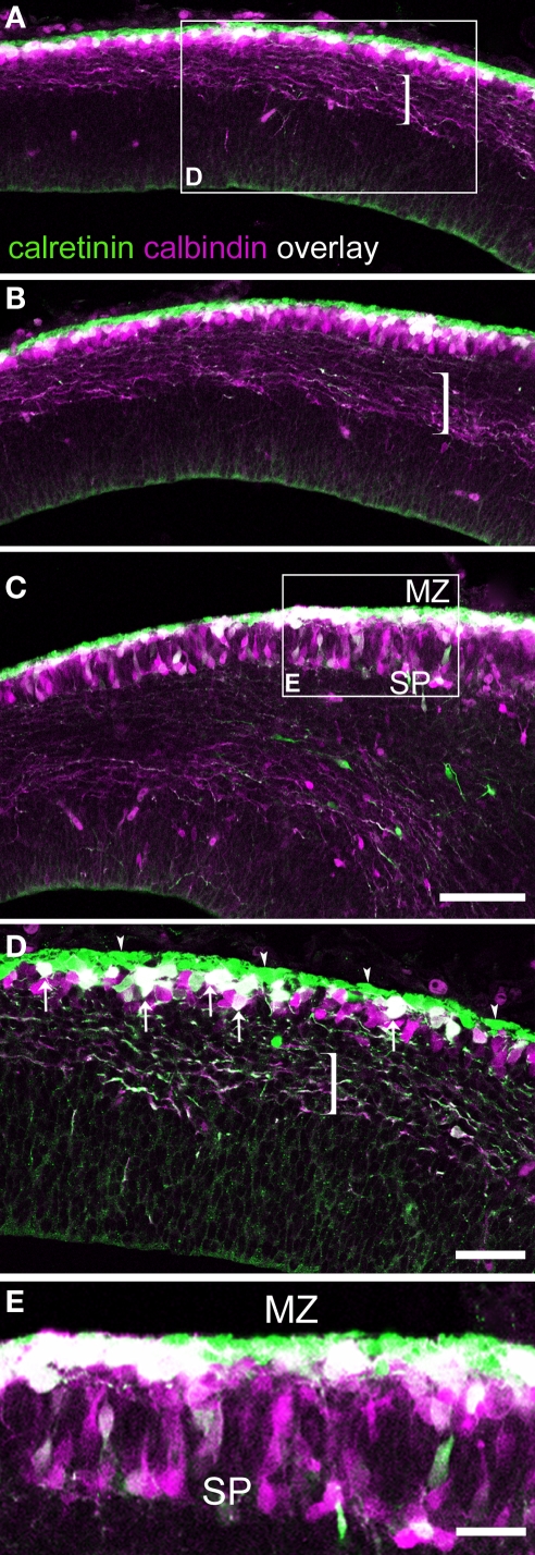
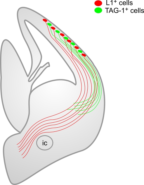
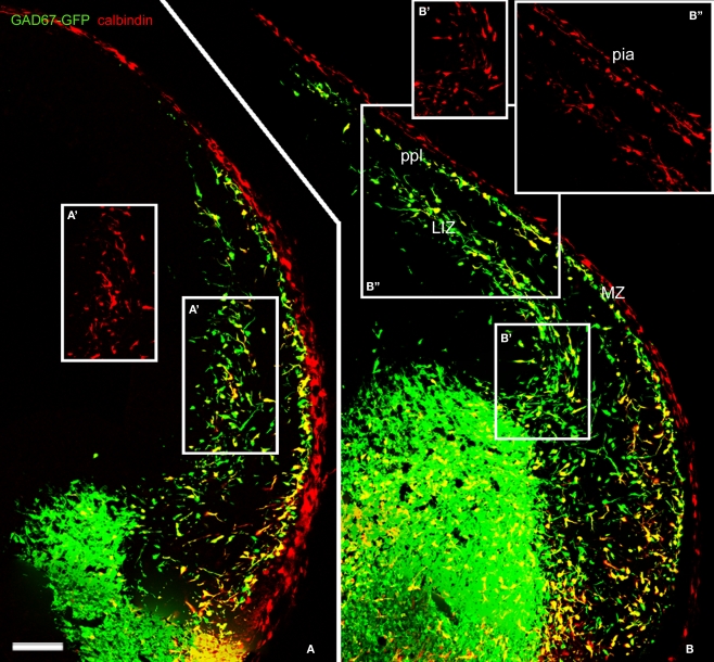
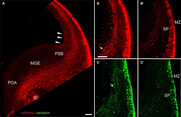
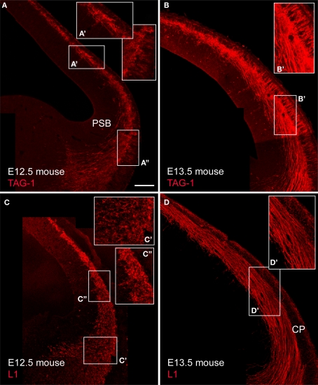
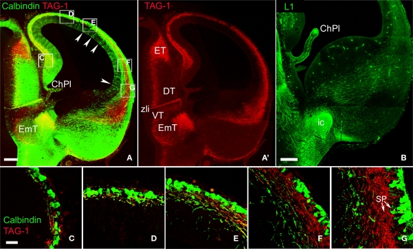
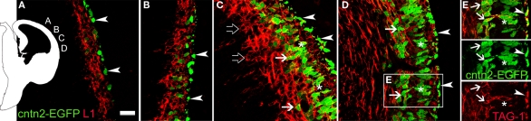
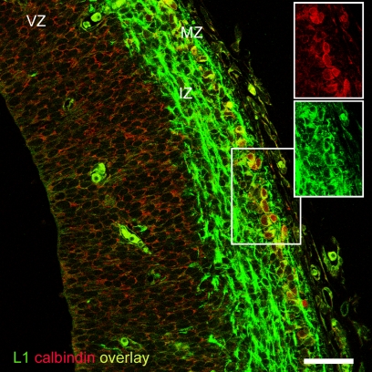
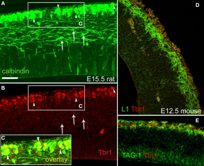
The preplate of the cerebral cortex contains projection neurons that connect the cortical primordium with the subpallium. These are collectively named pioneer neurons. After preplate partition, most of these pioneer neurons become subplate neurons. Certain preplate neurons, however, never associate with the subplate but rather with the marginal zone. In the present overview, we propose a novel classification of non-subplate pioneer neurons in rodents into two subtypes. In rats, the neurons of the first subtype are calbindin(+) (CB), calretinin(+) (CR) and L1(+) and are situated in the upper part of the preplate before its partition. Neurons of the second subtype are TAG-1(+) and are located slightly deeper to the previous population in the preplate. After the preplate partition, the CB(+), CR(+) and L1(+) neurons remain in the marginal zone whereas TAG-1(+) neurons become transiently localized in the upper cortical plate. In mice, by contrast, calcium binding proteins did not label pioneer neurons. We define in mice two subtypes of non-subplate pioneer neurons, either L1(+) or TAG-1(+)/cntn2(+). We propose these to be the homologues of the two subtypes of non-subplate pioneer neurons of rats. The anatomical distribution of these neuron populations is similar in rats and mice. The two populations of non-subplate pioneer neurons differ in their axonal projections. Axons of L1(+) pioneer neurons project to the ganglionic eminences and the anterior preoptic area, but avoid entering the posterior limb of the internal capsule towards the thalamus. Axons of TAG-1(+) pioneer neurons project to the lateral parts of the ganglionic eminences at the early stages of cortical histogenesis examined.
大脑皮层的基板含有投射神经元,这些神经元将皮质原基与皮质下区连接起来。这些神经元统称为先驱神经元。在基板分裂后,这些先驱神经元中的大多数成为基板下神经元。然而,某些基板前神经元从未与基板下区联系,而是与边缘区联系。在本综述中,我们提出了一种新的分类方法,将啮齿动物中非基板先驱神经元分为两种亚型。在大鼠中,第一种亚型的神经元是 calbindin(+) (CB)、calretinin(+) (CR) 和 L1(+),它们位于基板分裂前的基板上层。第二种亚型的神经元是 TAG-1(+),位于基板前区稍深的位置。基板分裂后,CB(+)、CR(+) 和 L1(+)神经元仍位于边缘区,而 TAG-1(+)神经元则暂时定位于上皮质板。相比之下,在小鼠中,钙结合蛋白没有标记先驱神经元。我们在小鼠中定义了两种非基板先驱神经元亚型,即 L1(+)或 TAG-1(+)/cntn2(+)。我们提出这些是大鼠两种非基板先驱神经元亚型的同源物。这些神经元群体的解剖分布在大鼠和小鼠中相似。两种非基板先驱神经元群体的轴突投射不同。L1(+)先驱神经元的轴突投射到神经节隆起和前视前区,但避免进入内侧囊后肢向丘脑。TAG-1(+)先驱神经元的轴突在皮质发生早期投射到神经节隆起的外侧部分。