Department of General Pathology, Institute of Biological Sciences, Federal University of Minas Gerais (UFMG), Belo Horizonte, Brazil.
Evid Based Complement Alternat Med. 2011;2011:182703. doi: 10.1093/ecam/nep197. Epub 2011 Mar 9.
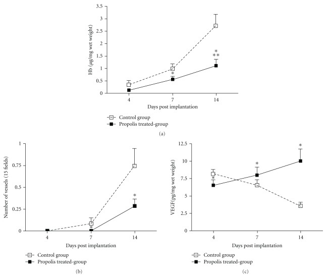
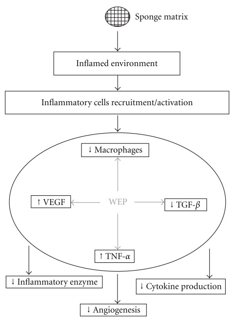
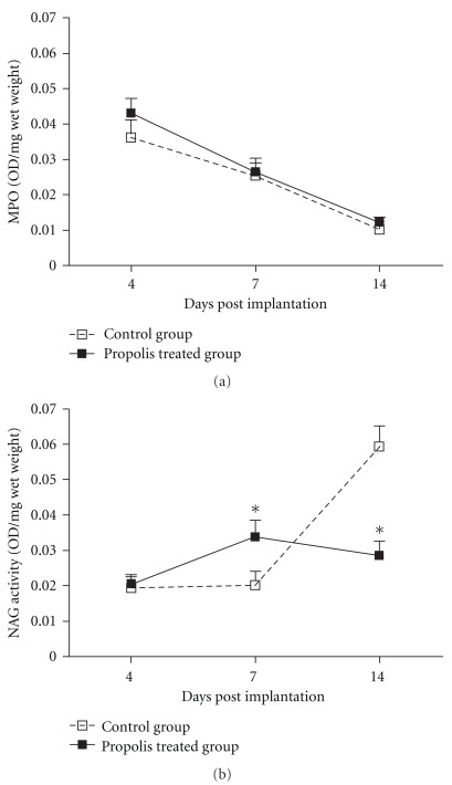
Angiogenesis and inflammation are persistent features of several pathological conditions. Propolis, a sticky material that honeybees collect from living plants, has been reported to have multiple biological effects including anti-inflammatory and anti-neoplasic activities. Here, we investigated the effects of water extract of green propolis (WEP) on angiogenesis, inflammatory cell accumulation and endogenous production of cytokines in sponge implants of mice over a 14-day period. Blood vessel formation as assessed by hemoglobin content and by morphometric analysis of the implants was reduced by WEP (500 mg kg(-1) orally) compared to the untreated group. The levels of vascular endothelial growth factor (VEGF) increased progressively in the treated group but decreased after Day 10 in the control group. Accumulation of neutrophils and macrophages was determined by measuring myeloperoxidase (MPO) and N-acetyl-β-(D)-glucosaminidase (NAG) activities, respectively. Neutrophil accumulation was unaffected by propolis, but NAG activity was reduced by the treatment at Day 14. The levels TGF-β1 intra-implant increased progressively in both groups but were higher (40%) at Day 14 in the control implants. The pro-inflammatory levels of TNF-α peaked at Day 7 in the control implants, and at Day 14 in the propolis-treated group. Our results indicate that the anti-inflammatory/anti-angiogenic effects of propolis are associated with cytokine modulation.
血管生成和炎症是几种病理状况的持续特征。蜂胶是蜜蜂从活植物中采集的粘性物质,据报道具有多种生物学效应,包括抗炎和抗瘤活性。在这里,我们研究了水提绿蜂胶(WEP)在 14 天期间对小鼠海绵植入物中的血管生成、炎性细胞积累和内源性细胞因子产生的影响。与未处理组相比,WEP(口服 500mg/kg)降低了血红蛋白含量和植入物形态计量分析评估的血管生成。在治疗组中,血管内皮生长因子(VEGF)的水平逐渐增加,但在对照组中第 10 天后下降。通过测量髓过氧化物酶(MPO)和 N-乙酰-β-(D)-氨基葡萄糖苷酶(NAG)活性分别确定中性粒细胞和巨噬细胞的积累。蜂胶对中性粒细胞的积累没有影响,但在第 14 天治疗时 NAG 活性降低。两组 TGF-β1 水平在植入物内均逐渐增加,但在对照组植入物中第 14 天更高(40%)。对照植入物中 TNF-α的促炎水平在第 7 天达到峰值,而在蜂胶治疗组中在第 14 天达到峰值。我们的结果表明,蜂胶的抗炎/抗血管生成作用与细胞因子调节有关。