Department of Comparative Medicine, Stanford University, California 94305, USA.
J Comp Neurol. 2010 Mar 1;518(5):647-67. doi: 10.1002/cne.22235.
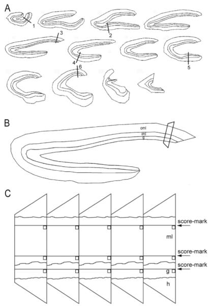
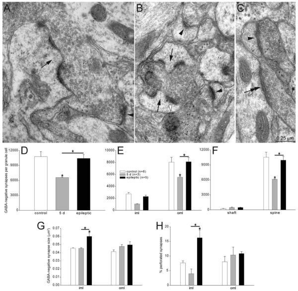
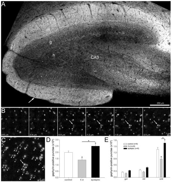
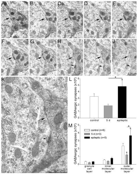
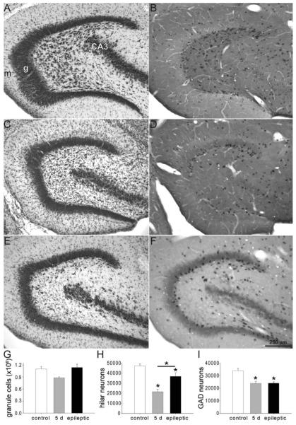
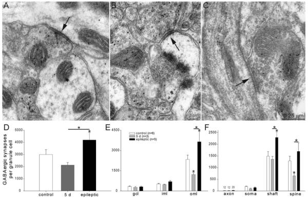
Many patients with temporal lobe epilepsy display neuron loss in the dentate gyrus. One potential epileptogenic mechanism is loss of GABAergic interneurons and inhibitory synapses with granule cells. Stereological techniques were used to estimate numbers of gephyrin-positive punctae in the dentate gyrus, which were reduced short-term (5 days after pilocarpine-induced status epilepticus) but later rebounded beyond controls in epileptic rats. Stereological techniques were used to estimate numbers of synapses in electron micrographs of serial sections processed for postembedding GABA-immunoreactivity. Adjacent sections were used to estimate numbers of granule cells and glutamic acid decarboxylase-positive neurons per dentate gyrus. GABAergic neurons were reduced to 70% of control levels short-term, where they remained in epileptic rats. Integrating synapse and cell counts yielded average numbers of GABAergic synapses per granule cell, which decreased short-term and rebounded in epileptic animals beyond control levels. Axo-shaft and axo-spinous GABAergic synapse numbers in the outer molecular layer changed most. These findings suggest interneuron loss initially reduces numbers of GABAergic synapses with granule cells, but later, synaptogenesis by surviving interneurons overshoots control levels. In contrast, the average number of excitatory synapses per granule cell decreased short-term but recovered only toward control levels, although in epileptic rats excitatory synapses in the inner molecular layer were larger than in controls. These findings reveal a relative excess of GABAergic synapses and suggest that reports of reduced functional inhibitory synaptic input to granule cells in epilepsy might be attributable not to fewer but instead to abundant but dysfunctional GABAergic synapses.
许多颞叶癫痫患者表现出齿状回神经元丢失。一种潜在的致痫机制是 GABA 能中间神经元和与颗粒细胞的抑制性突触丢失。立体学技术用于估计齿状回中 gephyrin 阳性斑点的数量,这些数量在 pilocarpine 诱导的癫痫持续状态后 5 天(短期)减少,但在癫痫大鼠中后来反弹超过对照。立体学技术用于估计 GABA 免疫反应性连续切片电子显微镜照片中突触的数量。相邻切片用于估计每个齿状回的颗粒细胞和谷氨酸脱羧酶阳性神经元的数量。GABA 能神经元在短期减少到对照水平的 70%,在癫痫大鼠中仍然如此。整合突触和细胞计数得出每个颗粒细胞的 GABA 能突触的平均数量,这些突触在癫痫动物中短期减少并反弹超过对照水平。外分子层的轴突-轴突棘 GABA 能突触数量变化最大。这些发现表明,中间神经元丢失最初会减少与颗粒细胞的 GABA 能突触数量,但随后存活的中间神经元的突触发生过度超过对照水平。相比之下,每个颗粒细胞的兴奋性突触的平均数量在短期减少,但仅恢复到对照水平,尽管在癫痫大鼠中内分子层的兴奋性突触比对照大鼠大。这些发现揭示了 GABA 能突触的相对过剩,并表明癫痫中颗粒细胞功能抑制性突触传入减少的报道可能不是由于突触数量减少,而是由于大量但功能失调的 GABA 能突触。