Iwata M, Sato A
Second Department of Internal Medicine, Hamamatsu University School of Medicine, Japan.
Infect Immun. 1991 Apr;59(4):1514-20. doi: 10.1128/iai.59.4.1514-1520.1991.
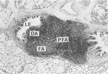
Pseudomonas aeruginosa is one of the most frequently encountered bacterial pathogens in patients with chronic pulmonary infections, including cystic fibrosis and diffuse panbronchiolitis. Bronchus-associated lymphoid tissue (BALT), noted frequently in patients with cystic fibrosis and diffuse panbronchiolitis, is considered to play an important role in the local immunologic defense mechanisms in the respiratory tract. To investigate the role of BALT in chronic pulmonary infections, we developed an animal model for chronic pulmonary infection and studied the morphological and immunohistochemical characteristics of BALT. Experimental pneumonia was produced in rats by intratracheal inoculation of P. aeruginosa enmeshed in agar beads. The histological changes corresponded to those occurring in chronic bronchiolitis. Immunohistochemically, surface immunoglobulin M-positive (sIgM+) cells and sIgA+ cells were recognized in the inflamed bronchial walls from day 4, and sIgG+ cells were recognized from day 14, W3/25+ cells exceeded OX8+ cells in number until day 14. In the BALT, there was a massive accumulation of lymphocytes in the lymphatics and high endothelial venules. The development of germinal centers was accompanied by increased numbers of sIgM+ and sIgA+ cells. W3/25+ cells exceeded OX8+ cells in number in the BALT until day 14. On the other hand, OX8+ cells were predominant in comparison with W3/25+ cells at day 21, and then both sIgM+ and sIgA+ cells and inflammatory changes in the lung decreased at day 28. These findings suggest that BALT regulates the local immune responses against chronic pulmonary infection due to P. aeruginosa.
铜绿假单胞菌是慢性肺部感染患者中最常见的细菌病原体之一,这些感染包括囊性纤维化和弥漫性泛细支气管炎。支气管相关淋巴组织(BALT)在囊性纤维化和弥漫性泛细支气管炎患者中经常可见,被认为在呼吸道局部免疫防御机制中发挥重要作用。为了研究BALT在慢性肺部感染中的作用,我们建立了一种慢性肺部感染动物模型,并研究了BALT的形态学和免疫组织化学特征。通过气管内接种包裹在琼脂珠中的铜绿假单胞菌在大鼠中诱发实验性肺炎。组织学变化与慢性支气管炎中发生的变化一致。免疫组织化学显示,从第4天起在炎症支气管壁中可识别出表面免疫球蛋白M阳性(sIgM+)细胞和sIgA+细胞,从第14天起可识别出sIgG+细胞,直到第14天W3/25+细胞数量超过OX8+细胞。在BALT中,淋巴管和高内皮静脉中有大量淋巴细胞聚集。生发中心的形成伴随着sIgM+和sIgA+细胞数量的增加。直到第14天,BALT中W3/25+细胞数量超过OX8+细胞。另一方面,在第21天时OX8+细胞与W3/25+细胞相比占主导地位,然后在第28天时sIgM+和sIgA+细胞以及肺部炎症变化均减少。这些发现表明,BALT调节针对铜绿假单胞菌引起的慢性肺部感染的局部免疫反应。