Redenti Stephen, Tao Sarah, Yang Jing, Gu Ping, Klassen Henry, Saigal Sunita, Desai Tejal, Young Michael J
J Ocul Biol Dis Infor. 2008 Mar;1(1):19-29. doi: 10.1007/s12177-008-9005-3. Epub 2008 May 22.
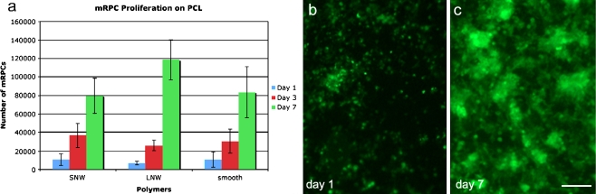
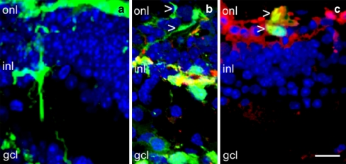
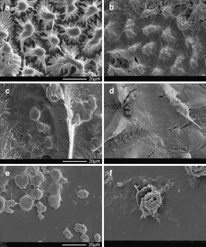
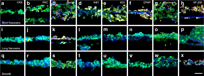
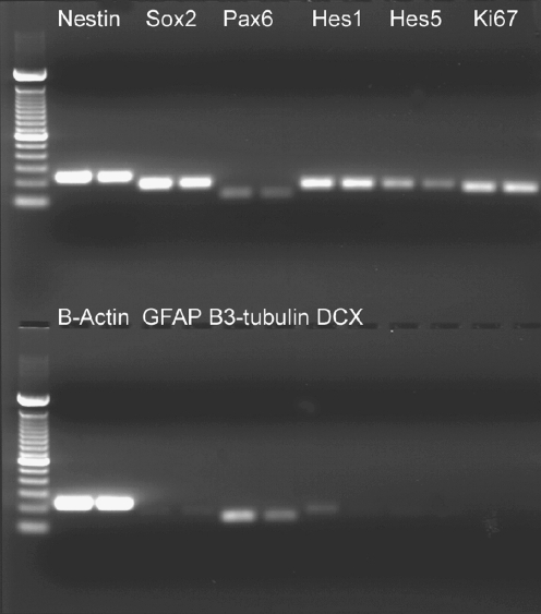
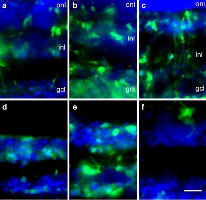
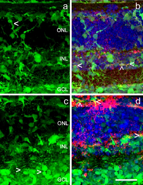
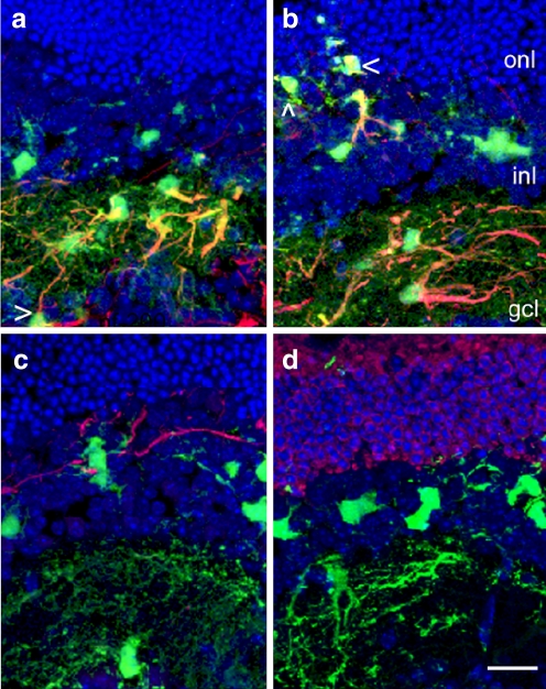
Retinal progenitor cells (RPCs) can be combined with nanostructured polymer scaffolds to generate composite grafts in culture. One strategy for repair of diseased retinal tissue involves implantation of composite grafts of this type in the subretinal space. In the present study, mouse retinal progenitor cells (RPCs) were cultured on laminin-coated novel nanowire poly(e-caprolactone)(PCL) scaffolds, and the survival, differentiation, and migration of these cells into the retina of C57bl/6 and rhodospsin -/- mouse retinal explants and transplant recipients were analyzed. RPCs were cultured on smooth PCL and both short (2.5 mum) and long (27 mum) nanowire PCL scaffolds. Scaffolds with adherent mRPCs were then either co-cultured with, or transplanted to, wild-type and rhodopsin -/- mouse retina. Robust RPC proliferation on each type of PCL scaffold was observed. Immunohistochemistry revealed that RPCs cultured on nanowire scaffolds increased expression of mature bipolar and photoreceptor markers. Reverse transcription polymerase chain reaction revealed down-regulation of several early progenitor markers. PCL-delivered RPCs migrated into the retina of both wild-type and rhodopsin knockout mice. The results provide evidence that RPCs proliferate and express mature retinal proteins in response to interactions with nanowire scaffolds. These composite grafts allow for the migration and differentiation of new cells into normal and degenerated retina.
视网膜祖细胞(RPCs)可与纳米结构聚合物支架相结合,在培养中生成复合移植物。修复病变视网膜组织的一种策略是将这种类型的复合移植物植入视网膜下间隙。在本研究中,将小鼠视网膜祖细胞(RPCs)培养在层粘连蛋白包被的新型纳米线聚己内酯(PCL)支架上,并分析这些细胞在C57bl/6和视紫红质基因敲除小鼠视网膜外植体及移植受体视网膜中的存活、分化和迁移情况。RPCs在光滑PCL以及短(2.5μm)和长(27μm)纳米线PCL支架上进行培养。然后将附着有mRPCs的支架与野生型和视紫红质基因敲除小鼠视网膜共同培养或移植到其视网膜中。观察到RPCs在每种类型的PCL支架上均有强劲增殖。免疫组织化学显示,在纳米线支架上培养的RPCs增加了成熟双极细胞和光感受器标志物的表达。逆转录聚合酶链反应显示几种早期祖细胞标志物表达下调。PCL递送的RPCs迁移到野生型和视紫红质基因敲除小鼠的视网膜中。结果表明,RPCs在与纳米线支架相互作用时会增殖并表达成熟的视网膜蛋白。这些复合移植物可使新细胞迁移并分化到正常和退化的视网膜中。