Department of Biomedical Engineering, Washington University, St Louis, MO 63130, USA.
Circ Res. 2010 Mar 19;106(5):981-91. doi: 10.1161/CIRCRESAHA.109.204891. Epub 2010 Jan 21.
Transmural dispersion of repolarization has been shown to play a role in the genesis of ventricular tachycardia and fibrillation in different animal models of heart failure (HF). Heterogeneous changes of repolarization within the midmyocardial population of ventricular cells have been considered an important contributor to the HF phenotype. However, there is limited electrophysiological data from the human heart.
To study electrophysiological remodeling of transmural repolarization in the failing and nonfailing human hearts.
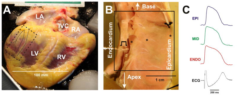
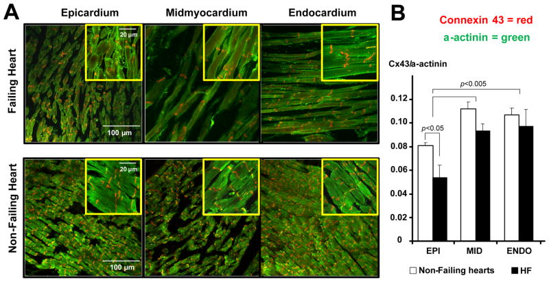
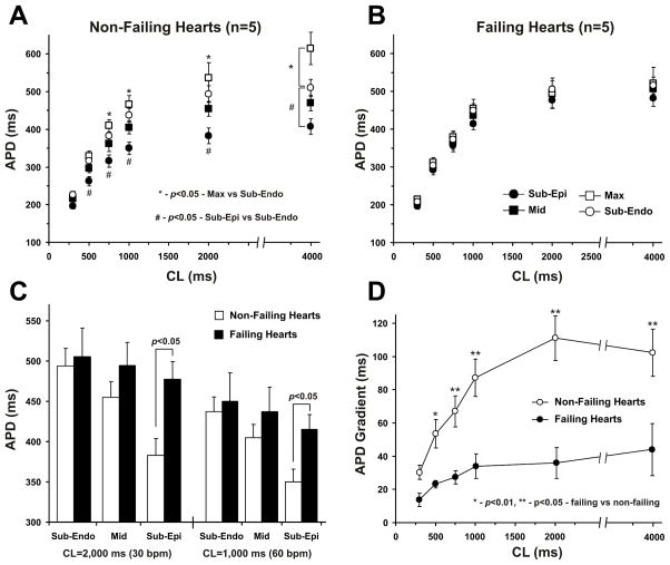
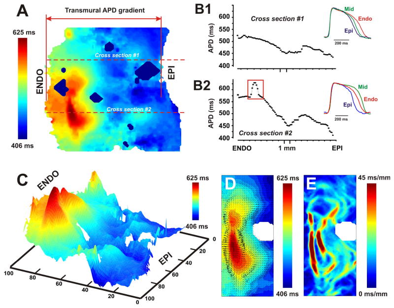
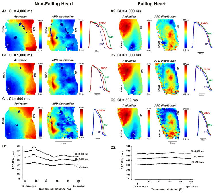
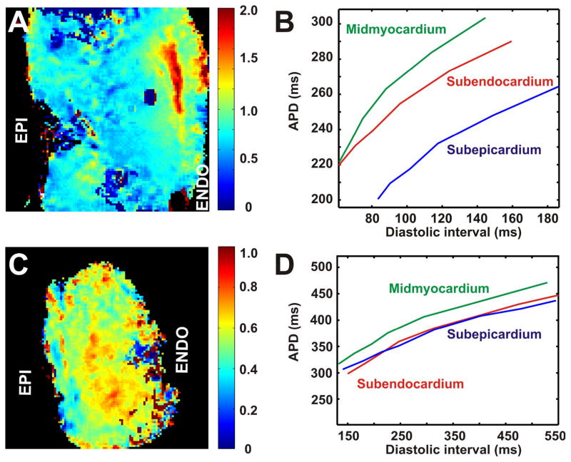
We optically mapped the action potential duration (APD) in the coronary-perfused scar-free posterior-lateral left ventricular free wall wedge preparations from failing (n=5) and nonfailing (n=5) human hearts. During slow pacing (S1S1=2000 ms), in the nonfailing hearts we observed significant transmural APD gradient: subepicardial, midmyocardial, and subendocardial APD80 were 383+/-21, 455+/-20, and 494+/-22 ms, respectively. In 60% of nonfailing hearts (3 of 5), we found midmyocardial islands of cells that presented a distinctly long APD (537+/-40 ms) and a steep local APD gradient (27+/-7 ms/mm) compared with the neighboring myocardium. HF resulted in prolongation of APD80: 477+/-22 ms, 495+/-29 ms, and 506+/-35 ms for the subepi-, mid-, and subendocardium, respectively, while reducing transmural APD80 difference from 111+/-13 to 29+/-6 ms (P<0.005) and presence of any prominent local APD gradient. In HF, immunostaining revealed a significant reduction of connexin43 expression on the subepicardium.
We present for the first time direct experimental evidence of a transmural APD gradient in the human heart. HF results in the heterogeneous prolongation of APD, which significantly reduces the transmural and local APD gradients.
已有研究表明,跨壁复极离散度在不同心力衰竭(HF)动物模型的室性心动过速和颤动的发生中发挥作用。心室细胞中层心肌内复极的异质性变化被认为是 HF 表型的一个重要因素。然而,来自人心的电生理数据有限。
研究衰竭和非衰竭人心的跨壁复极电重构。
我们在冠状灌注的无瘢痕左室游离壁后外侧楔形标本中光学标测动作电位时程(APD),标本来自衰竭(n=5)和非衰竭(n=5)人心。在缓慢起搏(S1S1=2000 ms)时,在非衰竭心脏中我们观察到明显的跨壁 APD 梯度:心外膜下、中层和心内膜下 APD80 分别为 383±21、455±20 和 494±22 ms。在 60%的非衰竭心脏(5 个中的 3 个)中,我们发现中层存在心肌小岛,其 APD 明显延长(537±40 ms),局部 APD 梯度陡峭(27±7 ms/mm),与邻近心肌相比。HF 导致 APD80 延长:心外膜下、中层和心内膜下分别为 477±22、495±29 和 506±35 ms,同时减少跨壁 APD80 差值从 111±13 到 29±6 ms(P<0.005)和任何明显的局部 APD 梯度的出现。在 HF 中,免疫染色显示心外膜下的连接蛋白 43 表达明显减少。
我们首次在人心提供了跨壁 APD 梯度的直接实验证据。HF 导致 APD 的异质延长,这显著降低了跨壁和局部 APD 梯度。