• 文献检索
  • 文档翻译
  • 深度研究
  • 学术资讯
  • Suppr Zotero 插件Zotero 插件
  • 邀请有礼
  • 套餐&价格
  • 历史记录
应用&插件
Suppr Zotero 插件Zotero 插件浏览器插件Mac 客户端Windows 客户端微信小程序
定价
高级版会员购买积分包购买API积分包
服务
文献检索文档翻译深度研究API 文档MCP 服务
关于我们
关于 Suppr公司介绍联系我们用户协议隐私条款
关注我们

Suppr 超能文献

核心技术专利:CN118964589B侵权必究
粤ICP备2023148730 号-1Suppr @ 2026

文献检索

告别复杂PubMed语法,用中文像聊天一样搜索,搜遍4000万医学文献。AI智能推荐,让科研检索更轻松。

立即免费搜索

文件翻译

保留排版,准确专业,支持PDF/Word/PPT等文件格式,支持 12+语言互译。

免费翻译文档

深度研究

AI帮你快速写综述,25分钟生成高质量综述,智能提取关键信息,辅助科研写作。

立即免费体验

在神经突中对高密度标记的突触进行光学检测,并利用组合型多重荧光突触标记物进行连接性绘图。

On optical detection of densely labeled synapses in neuropil and mapping connectivity with combinatorially multiplexed fluorescent synaptic markers.

机构信息

Department of Statistics and Center for Theoretical Neuroscience, Columbia University, New York, New York, USA.

出版信息

PLoS One. 2010 Jan 22;5(1):e8853. doi: 10.1371/journal.pone.0008853.

DOI:10.1371/journal.pone.0008853
PMID:20107507
原文链接:https://pmc.ncbi.nlm.nih.gov/articles/PMC2809746/
Abstract

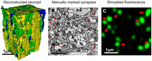
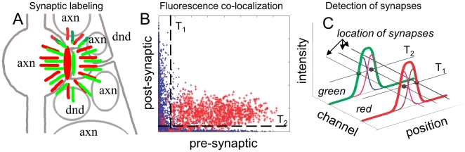
We propose a new method for mapping neural connectivity optically, by utilizing Cre/Lox system Brainbow to tag synapses of different neurons with random mixtures of different fluorophores, such as GFP, YFP, etc., and then detecting patterns of fluorophores at different synapses using light microscopy (LM). Such patterns will immediately report the pre- and post-synaptic cells at each synaptic connection, without tracing neural projections from individual synapses to corresponding cell bodies. We simulate fluorescence from a population of densely labeled synapses in a block of hippocampal neuropil, completely reconstructed from electron microscopy data, and show that high-end LM is able to detect such patterns with over 95% accuracy. We conclude, therefore, that with the described approach neural connectivity in macroscopically large neural circuits can be mapped with great accuracy, in scalable manner, using fast optical tools, and straightforward image processing. Relying on an electron microscopy dataset, we also derive and explicitly enumerate the conditions that should be met to allow synaptic connectivity studies with high-resolution optical tools.

摘要

我们提出了一种新的光学神经连接映射方法,利用 Cre/Lox 系统 Brainbow 将不同神经元的突触用不同荧光染料(如 GFP、YFP 等)的随机混合物进行标记,然后使用光学显微镜(LM)检测不同突触处的荧光模式。这种模式可以立即报告每个突触连接的前突触和后突触细胞,而无需从单个突触追踪神经投射到相应的细胞体。我们模拟了从电子显微镜数据完全重建的海马神经突块中高密度标记突触的荧光,并表明高端 LM 能够以超过 95%的准确率检测到这种模式。因此,我们得出结论,使用描述的方法,可以使用快速光学工具和简单的图像处理,以可扩展的方式,高精度地映射宏观上大型神经回路的神经连接。我们还依赖于一个电子显微镜数据集,推导出并明确列举了允许使用高分辨率光学工具进行突触连接研究的条件。

https://cdn.ncbi.nlm.nih.gov/pmc/blobs/46d2/2809746/55b0440687b8/pone.0008853.g007.jpg
https://cdn.ncbi.nlm.nih.gov/pmc/blobs/46d2/2809746/e5d8c57eb82e/pone.0008853.g001.jpg
https://cdn.ncbi.nlm.nih.gov/pmc/blobs/46d2/2809746/b66700f91360/pone.0008853.g002.jpg
https://cdn.ncbi.nlm.nih.gov/pmc/blobs/46d2/2809746/376dfac4b069/pone.0008853.g003.jpg
https://cdn.ncbi.nlm.nih.gov/pmc/blobs/46d2/2809746/d8421b7be6bc/pone.0008853.g004.jpg
https://cdn.ncbi.nlm.nih.gov/pmc/blobs/46d2/2809746/7bfa7fc2d5be/pone.0008853.g005.jpg
https://cdn.ncbi.nlm.nih.gov/pmc/blobs/46d2/2809746/997bafd6a072/pone.0008853.g006.jpg
https://cdn.ncbi.nlm.nih.gov/pmc/blobs/46d2/2809746/55b0440687b8/pone.0008853.g007.jpg

相似文献

1
On optical detection of densely labeled synapses in neuropil and mapping connectivity with combinatorially multiplexed fluorescent synaptic markers.在神经突中对高密度标记的突触进行光学检测,并利用组合型多重荧光突触标记物进行连接性绘图。
PLoS One. 2010 Jan 22;5(1):e8853. doi: 10.1371/journal.pone.0008853.
2
Advanced Fluorescence Protein-Based Synapse-Detectors.基于先进荧光蛋白的突触探测器
Front Synaptic Neurosci. 2016 Jun 30;8:16. doi: 10.3389/fnsyn.2016.00016. eCollection 2016.
3
Synaptic contacts between identified neurons visualized in the confocal laser scanning microscope. Neuroanatomical tracing combined with immunofluorescence detection of post-synaptic density proteins and target neuron-markers.在共聚焦激光扫描显微镜下观察到的特定神经元之间的突触联系。神经解剖学追踪与突触后密度蛋白和靶神经元标记物的免疫荧光检测相结合。
J Neurosci Methods. 2003 Sep 30;128(1-2):129-42. doi: 10.1016/s0165-0270(03)00171-7.
4
Reconstruction of complete connectivity matrix for connectomics by sampling neural connectivity with fluorescent synaptic markers.利用荧光突触标记物对神经连接进行采样,实现连接组学中完整连通性矩阵的重建。
J Neurosci Methods. 2011 Mar 30;196(2):289-302. doi: 10.1016/j.jneumeth.2011.01.021. Epub 2011 Jan 26.
5
3D structural imaging of the brain with photons and electrons.利用光子和电子对大脑进行三维结构成像。
Curr Opin Neurobiol. 2008 Dec;18(6):633-41. doi: 10.1016/j.conb.2009.03.005. Epub 2009 Apr 9.
6
A resource from 3D electron microscopy of hippocampal neuropil for user training and tool development.海马神经突 3D 电子显微镜资源,供用户培训和工具开发使用。
Sci Data. 2015 Sep 1;2:150046. doi: 10.1038/sdata.2015.46. eCollection 2015.
7
Volume electron microscopy of the distribution of synapses in the neuropil of the juvenile rat somatosensory cortex.少年大鼠感觉皮层神经胶内突触分布的电子显微镜体积研究。
Brain Struct Funct. 2018 Jan;223(1):77-90. doi: 10.1007/s00429-017-1470-7. Epub 2017 Jul 18.
8
Mapping the connectivity of serotonin transporter immunoreactive axons to excitatory and inhibitory neurochemical synapses in the mouse limbic brain.绘制小鼠边缘脑区中5-羟色胺转运体免疫反应性轴突与兴奋性和抑制性神经化学突触的连接图谱。
Brain Struct Funct. 2017 Apr;222(3):1297-1314. doi: 10.1007/s00429-016-1278-x. Epub 2016 Aug 2.
9
Tools and limitations to study the molecular composition of synapses by fluorescence microscopy.通过荧光显微镜研究突触分子组成的工具与局限性
Biochem J. 2016 Oct 15;473(20):3385-3399. doi: 10.1042/BCJ20160366.
10
Distribution and Structure of Synapses on Medial Vestibular Nuclear Neurons Targeted by Cerebellar Flocculus Purkinje Cells and Vestibular Nerve in Mice: Light and Electron Microscopy Studies.小鼠小脑绒球浦肯野细胞和前庭神经靶向的内侧前庭核神经元突触的分布与结构:光镜和电镜研究
PLoS One. 2016 Oct 6;11(10):e0164037. doi: 10.1371/journal.pone.0164037. eCollection 2016.

引用本文的文献

1
Quantitative Fluorescence Analysis Reveals Dendrite-Specific Thalamocortical Plasticity in L5 Pyramidal Neurons during Learning.定量荧光分析揭示了学习过程中 L5 锥体神经元树突特异性的丘脑皮层可塑性。
J Neurosci. 2023 Jan 25;43(4):584-600. doi: 10.1523/JNEUROSCI.1372-22.2022. Epub 2022 Dec 12.
2
Efficient and Accurate Synapse Detection With Selective Structured Illumination Microscopy on the Putative Regions of Interest of Ultrathin Serial Sections.在超薄连续切片的假定感兴趣区域上使用选择性结构光照显微镜进行高效准确的突触检测。
Front Neuroanat. 2021 Nov 15;15:759816. doi: 10.3389/fnana.2021.759816. eCollection 2021.
3

本文引用的文献

1
The structure of the nervous system of the nematode Caenorhabditis elegans.秀丽隐杆线虫的神经系统结构。
Philos Trans R Soc Lond B Biol Sci. 1986 Nov 12;314(1165):1-340. doi: 10.1098/rstb.1986.0056.
2
Semi-automated reconstruction of neural processes from large numbers of fluorescence images.从大量荧光图像中进行神经突起的半自动重建。
PLoS One. 2009 May 21;4(5):e5655. doi: 10.1371/journal.pone.0005655.
3
3D structural imaging of the brain with photons and electrons.利用光子和电子对大脑进行三维结构成像。
Gephyrin-Lacking PV Synapses on Neocortical Pyramidal Neurons.
缺少 Gephyrin 的 PV 突触在新皮层锥体神经元上。
Int J Mol Sci. 2021 Sep 17;22(18):10032. doi: 10.3390/ijms221810032.
4
Q&A: Array tomography.常见问题解答:面绘制技术
BMC Biol. 2018 Sep 6;16(1):98. doi: 10.1186/s12915-018-0560-1.
5
Using high-throughput barcode sequencing to efficiently map connectomes.利用高通量条形码测序技术高效绘制连接组图谱。
Nucleic Acids Res. 2017 Jul 7;45(12):e115. doi: 10.1093/nar/gkx292.
6
Synaptic circuitry of identified neurons in the antennal lobe of Drosophila melanogaster.黑腹果蝇触角叶中已鉴定神经元的突触回路。
J Comp Neurol. 2016 Jun 15;524(9):1920-56. doi: 10.1002/cne.23966. Epub 2016 Mar 9.
7
From a meso- to micro-scale connectome: array tomography and mGRASP.从中尺度到微观尺度的连接组学:阵列断层扫描和mGRASP。
Front Neuroanat. 2015 Jun 4;9:78. doi: 10.3389/fnana.2015.00078. eCollection 2015.
8
Designing tools for assumption-proof brain mapping.设计用于无假设脑图谱的工具。
Neuron. 2014 Sep 17;83(6):1239-41. doi: 10.1016/j.neuron.2014.09.004.
9
Thalamocortical input onto layer 5 pyramidal neurons measured using quantitative large-scale array tomography.使用定量大规模阵列断层摄影术测量丘脑皮层输入到 5 层锥体神经元。
Front Neural Circuits. 2013 Nov 12;7:177. doi: 10.3389/fncir.2013.00177. eCollection 2013.
10
Physical principles for scalable neural recording.可扩展神经记录的物理原理。
Front Comput Neurosci. 2013 Oct 21;7:137. doi: 10.3389/fncom.2013.00137. eCollection 2013.
Curr Opin Neurobiol. 2008 Dec;18(6):633-41. doi: 10.1016/j.conb.2009.03.005. Epub 2009 Apr 9.
4
A proposal for a coordinated effort for the determination of brainwide neuroanatomical connectivity in model organisms at a mesoscopic scale.关于在介观尺度上对模式生物全脑神经解剖连接性进行测定的协同努力的提议。
PLoS Comput Biol. 2009 Mar;5(3):e1000334. doi: 10.1371/journal.pcbi.1000334. Epub 2009 Mar 27.
5
The interscutularis muscle connectome.皮内肌连接组。
PLoS Biol. 2009 Feb 10;7(2):e32. doi: 10.1371/journal.pbio.1000032.
6
Automation of 3D reconstruction of neural tissue from large volume of conventional serial section transmission electron micrographs.从大量传统连续切片透射电子显微照片进行神经组织三维重建的自动化
J Neurosci Methods. 2009 Jan 30;176(2):276-89. doi: 10.1016/j.jneumeth.2008.09.006. Epub 2008 Sep 12.
7
Axon tracking in serial block-face scanning electron microscopy.连续块面扫描电子显微镜中的轴突追踪
Med Image Anal. 2009 Feb;13(1):180-8. doi: 10.1016/j.media.2008.05.002. Epub 2008 Jun 5.
8
Mapping the structural core of human cerebral cortex.绘制人类大脑皮层的结构核心。
PLoS Biol. 2008 Jul 1;6(7):e159. doi: 10.1371/journal.pbio.0060159.
9
A technicolour approach to the connectome.一种用于连接组的彩色方法。
Nat Rev Neurosci. 2008 Jun;9(6):417-22. doi: 10.1038/nrn2391. Epub 2008 Apr 30.
10
GFP Reconstitution Across Synaptic Partners (GRASP) defines cell contacts and synapses in living nervous systems.跨突触伙伴的绿色荧光蛋白重组(GRASP)可定义活体神经系统中的细胞接触和突触。
Neuron. 2008 Feb 7;57(3):353-63. doi: 10.1016/j.neuron.2007.11.030.