Lee Hojin, Oh Won Chan, Seong Jihye, Kim Jinhyun
Center for Functional Connectomics, Korea Institute of Science and TechnologySeoul, South Korea; Neuroscience Program, Korea University of Science and TechnologyDaejeon, South Korea.
Center for Functional Connectomics, Korea Institute of Science and Technology Seoul, South Korea.
Front Synaptic Neurosci. 2016 Jun 30;8:16. doi: 10.3389/fnsyn.2016.00016. eCollection 2016.
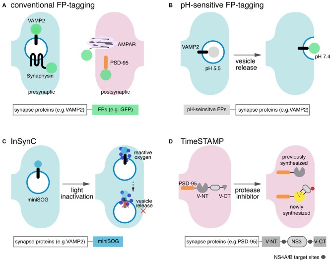
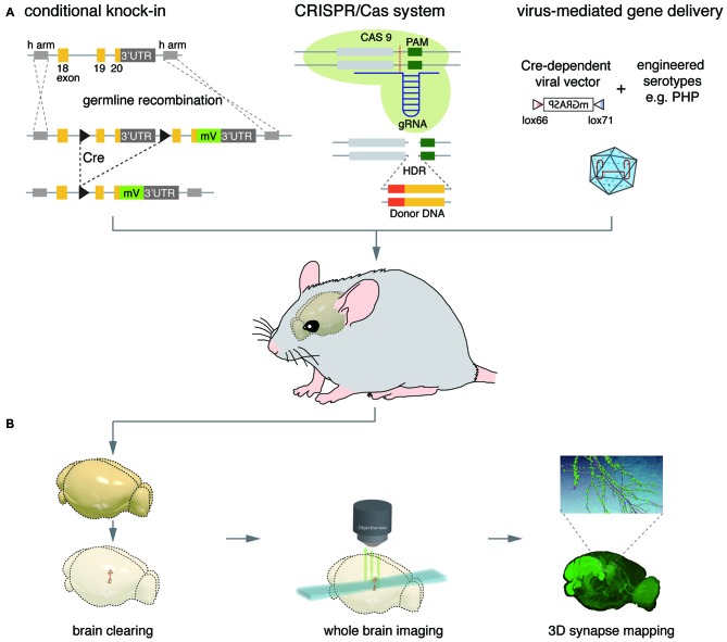
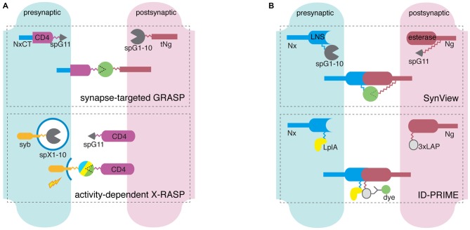
The complex information-processing capabilities of the central nervous system emerge from intricate patterns of synaptic input-output relationships among various neuronal circuit components. Understanding these capabilities thus requires a precise description of the individual synapses that comprise neural networks. Recent advances in fluorescent protein engineering, along with developments in light-favoring tissue clearing and optical imaging techniques, have rendered light microscopy (LM) a potent candidate for large-scale analyses of synapses, their properties, and their connectivity. Optically imaging newly engineered fluorescent proteins (FPs) tagged to synaptic proteins or microstructures enables the efficient, fine-resolution illumination of synaptic anatomy and function in large neural circuits. Here we review the latest progress in fluorescent protein-based molecular tools for imaging individual synapses and synaptic connectivity. We also identify associated technologies in gene delivery, tissue processing, and computational image analysis that will play a crucial role in bridging the gap between synapse- and system-level neuroscience.
中枢神经系统复杂的信息处理能力源自各种神经元回路组件之间错综复杂的突触输入-输出关系模式。因此,要理解这些能力,就需要精确描述构成神经网络的单个突触。荧光蛋白工程的最新进展,以及在光学透明组织和光学成像技术方面的发展,使光学显微镜(LM)成为大规模分析突触、其特性及其连接性的有力候选工具。对标记到突触蛋白或微观结构上的新工程化荧光蛋白(FPs)进行光学成像,能够在大型神经回路中对突触解剖结构和功能进行高效、高分辨率的照明。在此,我们综述了用于对单个突触和突触连接性进行成像的基于荧光蛋白的分子工具的最新进展。我们还确定了基因递送、组织处理和计算图像分析等相关技术,这些技术将在弥合突触水平和系统水平神经科学之间的差距方面发挥关键作用。