• 文献检索
  • 文档翻译
  • 深度研究
  • 学术资讯
  • Suppr Zotero 插件Zotero 插件
  • 邀请有礼
  • 套餐&价格
  • 历史记录
应用&插件
Suppr Zotero 插件Zotero 插件浏览器插件Mac 客户端Windows 客户端微信小程序
定价
高级版会员购买积分包购买API积分包
服务
文献检索文档翻译深度研究API 文档MCP 服务
关于我们
关于 Suppr公司介绍联系我们用户协议隐私条款
关注我们

Suppr 超能文献

核心技术专利:CN118964589B侵权必究
粤ICP备2023148730 号-1Suppr @ 2026

文献检索

告别复杂PubMed语法,用中文像聊天一样搜索,搜遍4000万医学文献。AI智能推荐,让科研检索更轻松。

立即免费搜索

文件翻译

保留排版,准确专业,支持PDF/Word/PPT等文件格式,支持 12+语言互译。

免费翻译文档

深度研究

AI帮你快速写综述,25分钟生成高质量综述,智能提取关键信息,辅助科研写作。

立即免费体验

免疫功能低下患者新型甲型H1N1流感感染的胸部CT表现。

Thoracic CT findings of novel influenza A (H1N1) infection in immunocompromised patients.

作者信息

Elicker Brett M, Schwartz Brian S, Liu Catherine, Chen Eunice C, Miller Steve A, Chiu Charles Y, Webb W Richard

机构信息

Department of Radiology and Biomedical Imaging, University of California, San Francisco, 505 Parnassus Avenue, P. O. Box 0628, San Francisco, CA, 94143, USA.

出版信息

Emerg Radiol. 2010 Jul;17(4):299-307. doi: 10.1007/s10140-010-0859-x. Epub 2010 Jan 29.

DOI:10.1007/s10140-010-0859-x
PMID:20111882
原文链接:https://pmc.ncbi.nlm.nih.gov/articles/PMC2880241/
Abstract

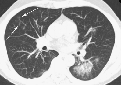
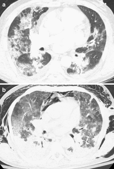
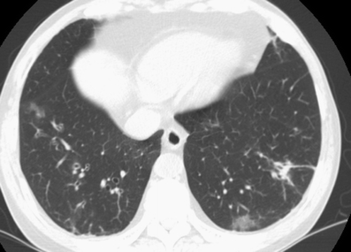
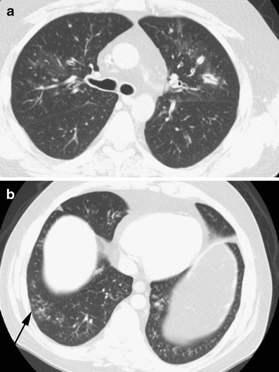
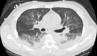
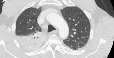
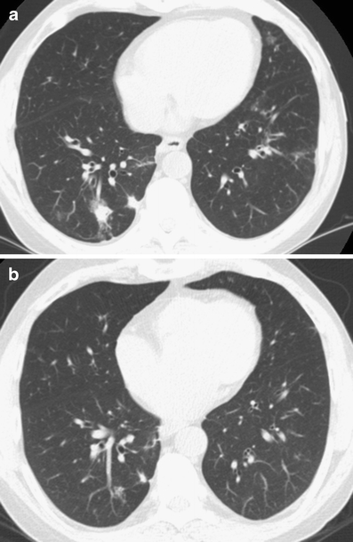
The goal of this study is to describe the spectrum of initial and follow-up CT findings of novel influenza A (H1N1) infection in a series of immunocompromised patients. Eight immunocompromised patients with documented novel influenza A (H1N1) had CT imaging at our institution between May 2009 and August 2009. A total of 20 CTs (initial and follow-up) were reviewed for the presence, severity, and distribution of the following: ground glass opacity, consolidation, interlobular septal thickening, mosaic perfusion, airway wall thickening, airway dilatation, nodules, cysts, pleural effusion, pericardial effusion, lymphadenopathy, and air trapping. The most common findings were airway thickening/dilatation, peribronchial ground glass opacity, centrilobular nodules, and tree-in-bud opacities. Peripheral consolidation involving the lower lobes was also a common pattern. Findings frequently involved all lobes and were closely associated with either large or small airways. Two patients presented with atypical CT findings including focal lobar consolidation and patchy lower lobe consolidation with soft tissue centrilobular nodules. Most survivors showed near complete resolution of findings within 35 days. CT scans in immunocompromised patients with novel influenza H1N1 commonly show a strong airway predominance of findings or peripheral areas of consolidation involving the lower lobes. A subset of patients with novel influenza A (H1N1) will show findings not typical of viral infection.

摘要

本研究的目的是描述一系列免疫功能低下患者新型甲型H1N1流感感染的初始及随访CT表现谱。2009年5月至2009年8月期间,8例确诊新型甲型H1N1流感的免疫功能低下患者在我们机构接受了CT检查。共回顾了20次CT扫描(初始及随访),以观察以下表现的存在、严重程度及分布情况:磨玻璃影、实变、小叶间隔增厚、马赛克灌注、气道壁增厚、气道扩张、结节、囊肿、胸腔积液、心包积液、淋巴结肿大及空气潴留。最常见的表现为气道增厚/扩张、支气管周围磨玻璃影、小叶中心结节及树芽征。累及下叶的外周实变也是常见表现形式。病变常累及所有肺叶,且与大小气道密切相关。2例患者出现非典型CT表现,包括局灶性肺叶实变及下叶斑片状实变伴软组织小叶中心结节。大多数幸存者在35天内病变几乎完全消退。免疫功能低下的新型甲型H1N1流感患者的CT扫描通常显示病变以气道为主或累及下叶的外周实变区域。一部分新型甲型H1N1流感患者会出现非典型病毒感染的表现。

https://cdn.ncbi.nlm.nih.gov/pmc/blobs/cb42/2880241/7ae47a683356/10140_2010_859_Fig8_HTML.jpg
https://cdn.ncbi.nlm.nih.gov/pmc/blobs/cb42/2880241/aeea654611a0/10140_2010_859_Fig1_HTML.jpg
https://cdn.ncbi.nlm.nih.gov/pmc/blobs/cb42/2880241/1ccf5a71e55d/10140_2010_859_Fig2_HTML.jpg
https://cdn.ncbi.nlm.nih.gov/pmc/blobs/cb42/2880241/c5cf1fa8c0b6/10140_2010_859_Fig3_HTML.jpg
https://cdn.ncbi.nlm.nih.gov/pmc/blobs/cb42/2880241/58079fa133bc/10140_2010_859_Fig4_HTML.jpg
https://cdn.ncbi.nlm.nih.gov/pmc/blobs/cb42/2880241/665d6a444ea8/10140_2010_859_Fig5_HTML.jpg
https://cdn.ncbi.nlm.nih.gov/pmc/blobs/cb42/2880241/22faef384c22/10140_2010_859_Fig6_HTML.jpg
https://cdn.ncbi.nlm.nih.gov/pmc/blobs/cb42/2880241/0ae7d6ba5e7b/10140_2010_859_Fig7_HTML.jpg
https://cdn.ncbi.nlm.nih.gov/pmc/blobs/cb42/2880241/7ae47a683356/10140_2010_859_Fig8_HTML.jpg

相似文献

1
Thoracic CT findings of novel influenza A (H1N1) infection in immunocompromised patients.免疫功能低下患者新型甲型H1N1流感感染的胸部CT表现。
Emerg Radiol. 2010 Jul;17(4):299-307. doi: 10.1007/s10140-010-0859-x. Epub 2010 Jan 29.
2
Novel influenza A (H1N1) virus infection in children: chest radiographic and CT evaluation.儿童新型甲型 H1N1 流感病毒感染:胸部 X 线摄影与 CT 评估。
Korean J Radiol. 2010 Nov-Dec;11(6):656-64. doi: 10.3348/kjr.2010.11.6.656. Epub 2010 Oct 29.
3
64 multidetector CT findings of influenza A (H1N1) virus in patients with hematologic malignancies.64例血液系统恶性肿瘤患者甲型H1N1流感病毒的多排CT表现
Acta Radiol. 2012 Jul;53(6):662-7. doi: 10.1258/ar.2012.120038. Epub 2012 Jun 25.
4
Swine-origin influenza A viral (H1N1) infection in children: chest computed tomography findings.儿童感染猪源甲型 H1N1 流感病毒:胸部计算机断层扫描结果。
Jpn J Radiol. 2011 Dec;29(10):707-11. doi: 10.1007/s11604-011-0619-1. Epub 2011 Oct 19.
5
Comparison of chest-CT findings of Influenza virus-associated pneumonia in immunocompetent vs. immunocompromised patients.免疫功能正常与免疫功能低下患者流感病毒相关性肺炎的胸部CT表现比较。
Eur J Radiol. 2015 Jun;84(6):1177-83. doi: 10.1016/j.ejrad.2015.02.014. Epub 2015 Feb 21.
6
High-resolution computed tomography findings of swine-origin influenza A (H1N1) virus (S-OIV) infection: comparison with scrub typhus.甲型H1N1流感病毒(S-OIV)感染的高分辨率计算机断层扫描结果:与恙虫病的比较。
Acta Radiol. 2012 Jul;53(6):657-61. doi: 10.1258/ar.2012.120012. Epub 2012 May 27.
7
Various computed tomography findings of 2009 H1N1 influenza in 17 patients with relatively mild illness.17 例轻症 2009H1N1 流感患者的各种计算机断层扫描表现。
Jpn J Radiol. 2011 Jun;29(5):301-6. doi: 10.1007/s11604-010-0558-2. Epub 2011 Jun 30.
8
Influenza A (H1N1) pneumonia: HRCT findings.甲型 H1N1 流感肺炎:HRCT 表现。
J Bras Pneumol. 2013 May-Jun;39(3):323-9. doi: 10.1590/S1806-37132013000300009.
9
Chest Radiological Findings of Patients With Severe H1N1 Pneumonia Requiring Intensive Care.需要重症监护的重症甲型H1N1肺炎患者的胸部放射学表现
J Intensive Care Med. 2016 Jan;31(1):51-60. doi: 10.1177/0885066614538753. Epub 2014 Jun 12.
10
H1N1 pneumonia: our experience in 50 patients with a severe clinical course of novel swine-origin influenza A (H1N1) virus (S-OIV).H1N1 肺炎:我们在 50 例新型猪源甲型 H1N1 流感病毒(S-OIV)所致严重临床病程患者中的经验。
Radiol Med. 2012 Mar;117(2):165-84. doi: 10.1007/s11547-011-0734-1. Epub 2011 Oct 21.

引用本文的文献

1
Investigation of high-resolution computed tomographic (HRCT) outcomes associated with chronic pulmonary microaspiration (CPM) in Tehran and Zahedan, Iran.伊朗德黑兰和扎黑丹与慢性肺部微吸入(CPM)相关的高分辨率计算机断层扫描(HRCT)结果研究。
Afr Health Sci. 2020 Dec;20(4):1710-1715. doi: 10.4314/ahs.v20i4.22.
2
Covid-19 imaging: A narrative review.新型冠状病毒肺炎影像学:一篇叙述性综述。
Ann Med Surg (Lond). 2021 Sep;69:102489. doi: 10.1016/j.amsu.2021.102489. Epub 2021 Jun 18.
3
COVID-19 or non-COVID viral pneumonia: How to differentiate based on the radiologic findings?

本文引用的文献

1
Swine-origin influenza A (H1N1) viral infection: radiographic and CT findings.猪源甲型 H1N1 流感病毒感染:影像学和 CT 表现。
AJR Am J Roentgenol. 2009 Dec;193(6):1494-9. doi: 10.2214/AJR.09.3625.
2
Chest radiographic and CT findings in novel swine-origin influenza A (H1N1) virus (S-OIV) infection.新型猪源甲型 H1N1 流感病毒(S-OIV)感染的胸部 X 线和 CT 表现。
AJR Am J Roentgenol. 2009 Dec;193(6):1488-93. doi: 10.2214/AJR.09.3599.
3
Critically Ill patients with 2009 influenza A(H1N1) in Mexico.墨西哥2009年甲型H1N1流感危重症患者。
新型冠状病毒肺炎(COVID-19)还是非新型冠状病毒病毒性肺炎:如何根据影像学表现进行鉴别?
World J Radiol. 2020 Dec 28;12(12):289-301. doi: 10.4329/wjr.v12.i12.289.
4
Vital role of chest CT in diagnosis of coronavirus disease 2019 (COVID-19).胸部CT在2019冠状病毒病(COVID-19)诊断中的重要作用。
Caspian J Intern Med. 2020 May;11(3):244-249. doi: 10.22088/cjim.11.3.244.
5
Chest Computed Tomography Findings in COVID-19 and Influenza: A Narrative Review.新型冠状病毒肺炎与流感的胸部计算机断层扫描表现:一项叙述性综述。
Biomed Res Int. 2020 Jun 5;2020:6928368. doi: 10.1155/2020/6928368. eCollection 2020.
6
Longitudinal changes of pneumonia complicating novel influenza A (H1N1) by high-resolution computed tomography.新型甲型H1N1流感并发肺炎的高分辨率计算机断层扫描的纵向变化
Radiol Infect Dis. 2015 Jun;2(1):40-46. doi: 10.1016/j.jrid.2015.06.001. Epub 2015 Jun 9.
7
Pulmonary High-Resolution Computed Tomography (HRCT) Findings of Patients with Early-Stage Coronavirus Disease 2019 (COVID-19) in Hangzhou, China.中国杭州早期 2019 冠状病毒病(COVID-19)患者的肺部高分辨率计算机断层扫描(HRCT)表现。
Med Sci Monit. 2020 Apr 4;26:e923885. doi: 10.12659/MSM.923885.
8
Community acquired respiratory virus infections in cancer patients-Guideline on diagnosis and management by the Infectious Diseases Working Party of the German Society for haematology and Medical Oncology.癌症患者社区获得性呼吸道病毒感染——德国血液学和医学肿瘤学会传染病工作组诊断与管理指南
Eur J Cancer. 2016 Nov;67:200-212. doi: 10.1016/j.ejca.2016.08.015. Epub 2016 Sep 25.
9
Lung in dengue: computed tomography findings.登革热肺部表现:计算机断层扫描表现。
PLoS One. 2014 May 16;9(5):e96313. doi: 10.1371/journal.pone.0096313. eCollection 2014.
10
Influenza A (H1N1) pneumonia: HRCT findings.甲型 H1N1 流感肺炎:HRCT 表现。
J Bras Pneumol. 2013 May-Jun;39(3):323-9. doi: 10.1590/S1806-37132013000300009.
JAMA. 2009 Nov 4;302(17):1880-7. doi: 10.1001/jama.2009.1536. Epub 2009 Oct 12.
4
Hospitalized patients with 2009 H1N1 influenza in the United States, April-June 2009.2009年4月至6月在美国住院治疗的2009年甲型H1N1流感患者。
N Engl J Med. 2009 Nov 12;361(20):1935-44. doi: 10.1056/NEJMoa0906695. Epub 2009 Oct 8.
5
Surveillance for the 2009 pandemic influenza A (H1N1) virus and seasonal influenza viruses - New Zealand, 2009.2009年新西兰甲型H1N1流感大流行病毒及季节性流感病毒监测
MMWR Morb Mortal Wkly Rep. 2009 Aug 28;58(33):918-21.
6
Epidemiological characteristics of pandemic influenza H1N1 2009 and seasonal influenza infection.2009年甲型H1N1大流行性流感和季节性流感感染的流行病学特征。
Med J Aust. 2009 Aug 3;191(3):146-9. doi: 10.5694/j.1326-5377.2009.tb02723.x.
7
H1N1 2009 influenza virus infection during pregnancy in the USA.美国孕期感染2009年甲型H1N1流感病毒的情况。
Lancet. 2009 Aug 8;374(9688):451-8. doi: 10.1016/S0140-6736(09)61304-0. Epub 2009 Jul 28.
8
Pneumonia and respiratory failure from swine-origin influenza A (H1N1) in Mexico.墨西哥出现的源自猪的甲型H1N1流感所致肺炎及呼吸衰竭
N Engl J Med. 2009 Aug 13;361(7):680-9. doi: 10.1056/NEJMoa0904252. Epub 2009 Jun 29.
9
The 2009 H1N1 influenza outbreak in its historical context.2009年甲型H1N1流感疫情的历史背景。
J Clin Virol. 2009 Jul;45(3):174-8. doi: 10.1016/j.jcv.2009.06.004. Epub 2009 Jun 11.
10
Chest CT findings of influenza virus-associated pneumonia in 12 adult patients.12例成年患者流感病毒相关性肺炎的胸部CT表现
Influenza Other Respir Viruses. 2007 Sep-Nov;1(5-6):183-7. doi: 10.1111/j.1750-2659.2008.00034.x.