McCarthy K P, Sloane J P, Kabarowski J H, Matutes E, Wiedemann L M
Leukaemia Research Fund Centre, Institute of Cancer Research, Chester Beatty Laboratories, London, United Kingdom.
Am J Pathol. 1991 Apr;138(4):821-8.
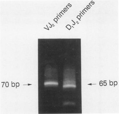
A series of T-cell proliferations in peripheral blood, bone marrow, or tissue samples were analyzed for clonality. The technique used employs the polymerase chain reaction to amplify portions of the rearranged T-cell receptor beta chain genes, using primers recognizing conserved sequences of the variable, diversity, and joining region segments. We examined 17 cases of T-cell lymphoma or leukemia; a clone was identified in 13 cases (76%) overall and in 7 of 8 cases (87.5%) in which both beta-chain alleles were known to be rearranged, as shown by restriction enzyme analysis. No clonal rearrangements were detected in samples from 13 non-T-cell disorders, including B-cell lymphomas, reactive lymphoid proliferations, and nonlymphoid tumors. This method is useful for detecting clones in thymic and post-thymic T-cell malignancies and has the advantages of being extremely rapid (a result is obtained within hours of the biopsy procedure), requiring no radiolabeling, using only a small amount of tissue, and being applicable to formalin-fixed, paraffin-embedded tissue.
对外周血、骨髓或组织样本中的一系列T细胞增殖进行克隆性分析。所采用的技术利用聚合酶链反应来扩增重排的T细胞受体β链基因的部分片段,使用识别可变区、多样区和连接区片段保守序列的引物。我们检测了17例T细胞淋巴瘤或白血病;总体上在13例(76%)中鉴定出克隆,在已知两条β链等位基因均发生重排的8例中的7例(87.5%)中鉴定出克隆,这通过限制性酶切分析得以显示。在13例非T细胞疾病的样本中未检测到克隆性重排,这些疾病包括B细胞淋巴瘤、反应性淋巴组织增生和非淋巴样肿瘤。该方法对于检测胸腺和胸腺后T细胞恶性肿瘤中的克隆很有用,并且具有极其快速(活检操作后数小时内即可获得结果)、无需放射性标记、仅需少量组织以及适用于福尔马林固定、石蜡包埋组织等优点。