Department of Cell Biology and Neuroscience, Montana State University, Bozeman, MT 59717, USA.
BMC Biotechnol. 2010 Feb 2;10:6. doi: 10.1186/1472-6750-10-6.
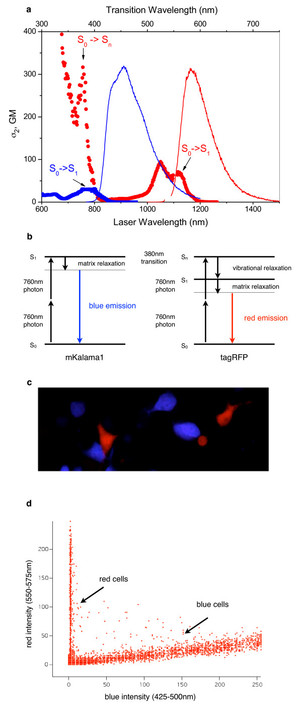
Two-photon dual-color imaging of tissues and cells labeled with fluorescent proteins (FPs) is challenging because most two-photon microscopes only provide one laser excitation wavelength at a time. At present, methods for two-photon dual-color imaging are limited due to the requirement of large differences in Stokes shifts between the FPs used and their low two-photon absorption (2PA) efficiency.
Here we present a new method of dual-color two-photon microscopy that uses the simultaneous excitation of the lowest-energy electronic transition of a blue fluorescent protein and a higher-energy electronic transition of a red fluorescent protein.
Our method does not require large differences in Stokes shifts and can be extended to a variety of FP pairs with larger 2PA efficiency and more optimal imaging properties.
用荧光蛋白(FPs)标记的组织和细胞的双光子双色成像具有挑战性,因为大多数双光子显微镜一次只能提供一个激光激发波长。目前,由于所用 FPs 的斯托克斯位移差异较大且双光子吸收(2PA)效率较低,双光子双色成像的方法受到限制。
本文提出了一种新的双光子显微镜双色成像方法,该方法同时激发蓝色荧光蛋白的最低能量电子跃迁和红色荧光蛋白的更高能量电子跃迁。
我们的方法不需要大的斯托克斯位移差异,可以扩展到具有更大 2PA 效率和更优成像特性的各种 FP 对。