Translational and Molecular Imaging Institute, Mount Sinai School of Medicine, Atran BM-24, Box 1234, One Gustave L. Levy Place, New York, NY 10029, USA.
Arterioscler Thromb Vasc Biol. 2010 Mar;30(3):403-10. doi: 10.1161/ATVBAHA.109.198556. Epub 2010 Feb 5.
Atherosclerotic plaque rupture leads to acute thrombus formation and may trigger serious clinical events such as myocardial infarction or stroke. Therefore, it would be valuable to identify atherothrombosis and vulnerable plaques before the onset of such clinical events. We sought to determine whether the noninvasive in vivo visualization of activated platelets was effective when using a target-specific MRI contrast agent to identify thrombi, hallmarks of vulnerable or high-risk atherosclerotic plaques.
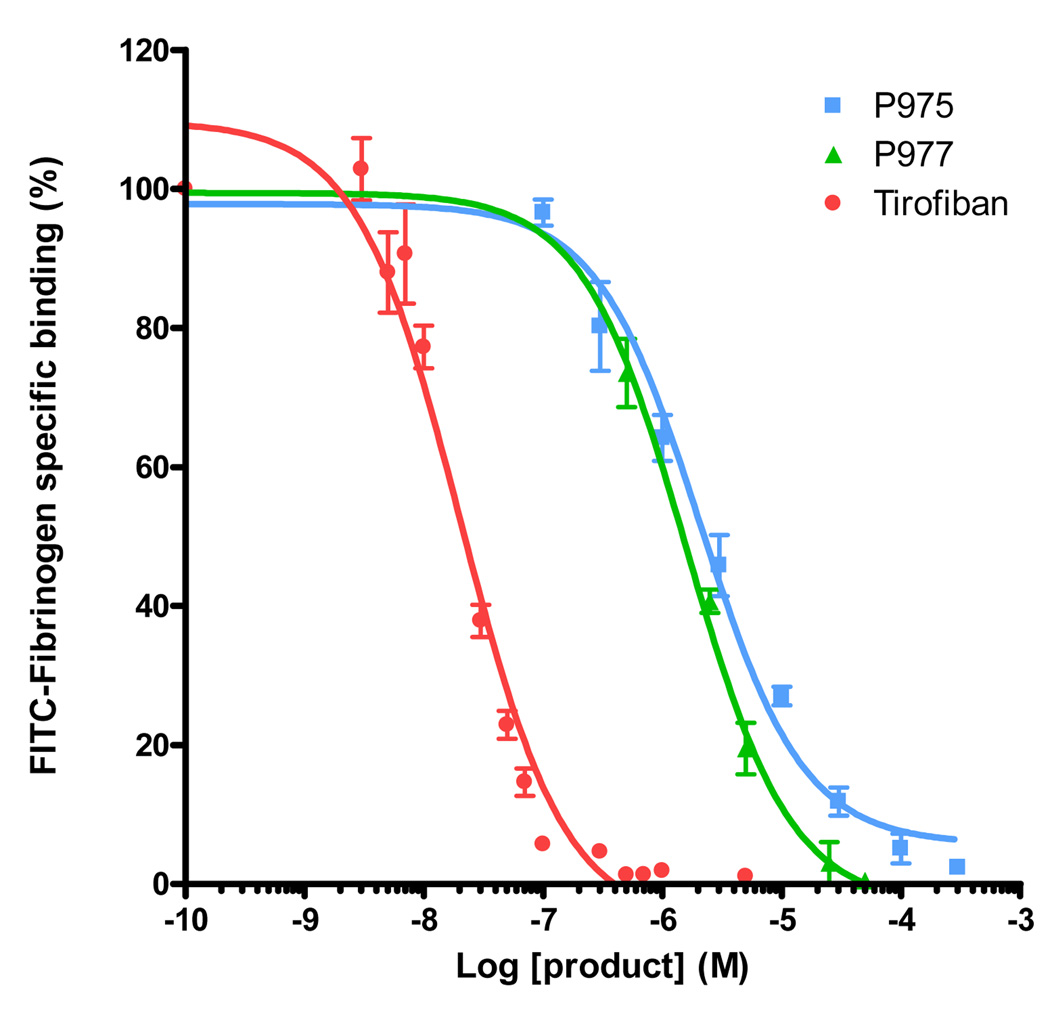
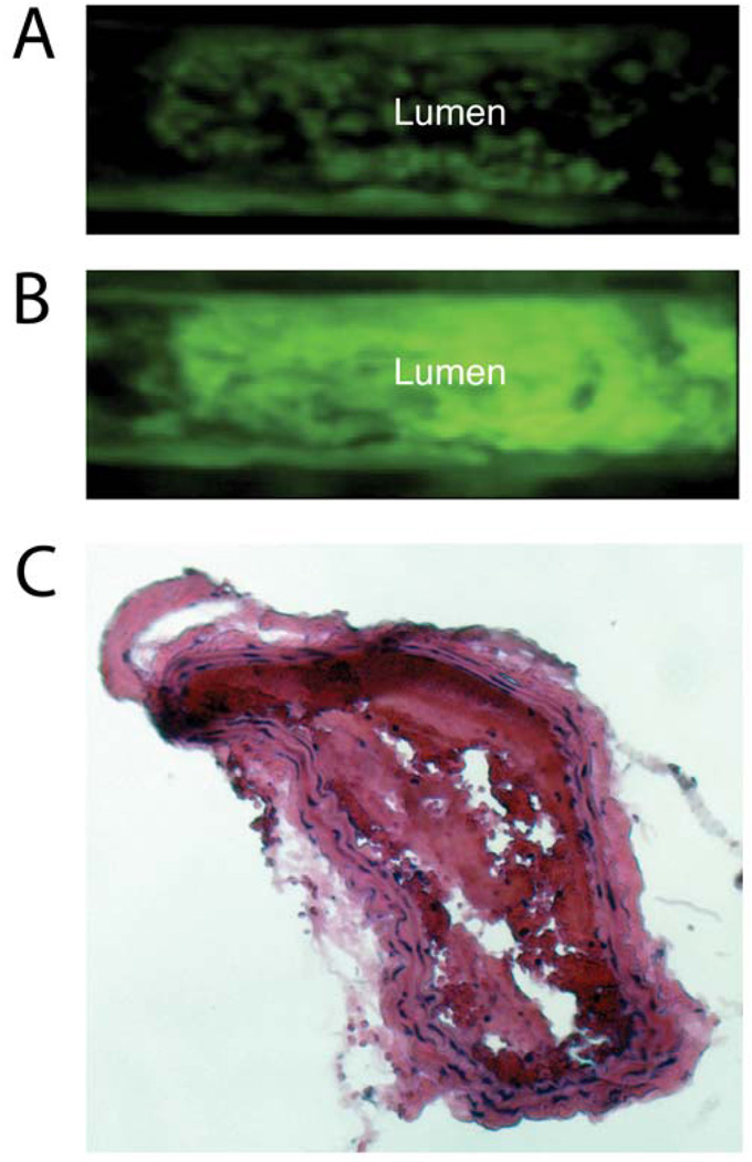
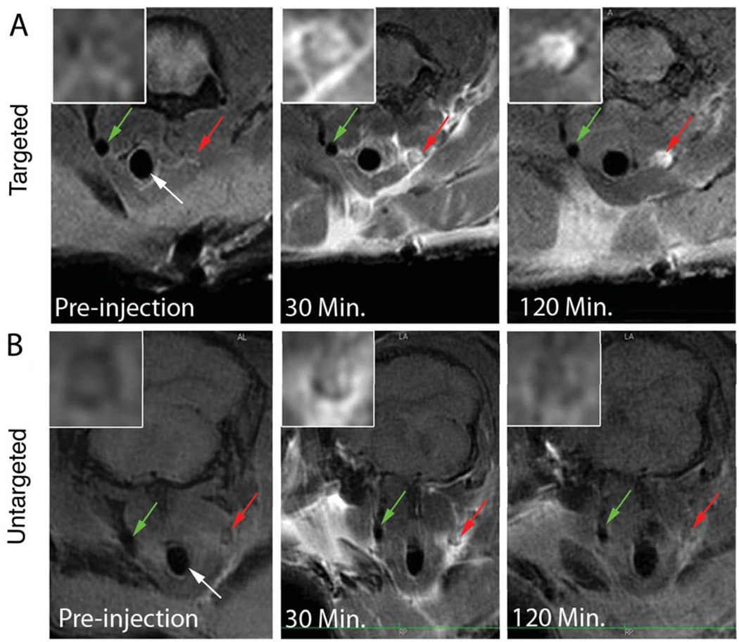
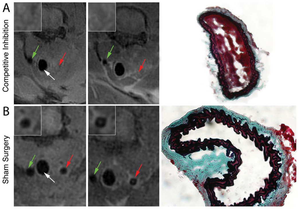
Inflammatory thrombi were induced in mice via topical application of arachidonic acid on the carotid. Thrombus formation was imaged with intravital fluorescence microscopy and molecular MRI. To accomplish the latter, a paramagnetic contrast agent (P975) that targets the glycoprotein alpha(IIb)beta(3), expressed on activated platelets, was investigated. The specificity of P975 for activated platelets was studied in vitro. In vivo, high spatial-resolution MRI was performed at baseline and longitudinally over 2 hours after injecting P975 or a nonspecific agent. The contralateral carotid, a sham surgery group, and a competitive inhibition experiment served as controls. P975 showed a good affinity for activated platelets, with an IC(50) (concentration of dose that produces 50% inhibition) value of 2.6 micromol/L. In thrombosed animals, P975 produced an immediate and sustained increase in MRI signal, whereas none of the control groups revealed any significant increase in MRI signal 2 hours after injection. More important, the competitive inhibition experiment with an alpha(IIb)beta(3) antagonist suppressed the MRI signal enhancement, which is indicative for the specificity of P975 for the activated platelets.
P975 allowed in vivo target-specific noninvasive MRI of activated platelets.
动脉粥样硬化斑块破裂可导致急性血栓形成,并可能引发严重的临床事件,如心肌梗死或中风。因此,在发生此类临床事件之前识别动脉血栓形成和易损斑块将具有重要价值。我们试图确定使用靶向特异性 MRI 造影剂识别血栓(易损或高危动脉粥样硬化斑块的标志物)时,体内激活血小板的非侵入性可视化是否有效。
通过在颈动脉表面应用花生四烯酸,在小鼠中诱导炎症性血栓形成。通过活体荧光显微镜和分子 MRI 对血栓形成进行成像。为了实现后者,研究了一种针对糖蛋白 α(IIb)β(3)的顺磁对比剂 (P975),该蛋白在激活的血小板上表达。在体外研究了 P975 对激活血小板的特异性。在体内,在注射 P975 或非特异性试剂后 2 小时内进行高空间分辨率 MRI。对侧颈动脉、假手术组和竞争性抑制实验作为对照。P975 对激活血小板具有良好的亲和力,IC(50)(产生 50%抑制的剂量浓度)值为 2.6 微摩尔/升。在血栓形成的动物中,P975 立即产生并持续增加 MRI 信号,而对照组在注射后 2 小时内均未显示出任何明显的 MRI 信号增加。更重要的是,用 α(IIb)β(3)拮抗剂进行的竞争性抑制实验抑制了 MRI 信号增强,这表明 P975 对激活血小板具有特异性。
P975 允许体内靶向特异性非侵入性 MRI 检测激活的血小板。