Proteobioactives Pty Ltd, 27/9 Powells Road, Brookvale, New South Wales 2100, Australia.
Arthritis Res Ther. 2010;12(1):R28. doi: 10.1186/ar2935. Epub 2010 Feb 18.
This study was undertaken to determine whether the anti-osteoarthritis drug pentosan polysulfate (PPS) influenced mesenchymal precursor cell (MPC) proliferation and differentiation.
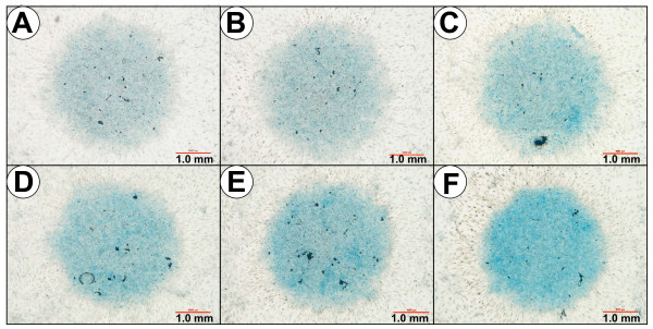
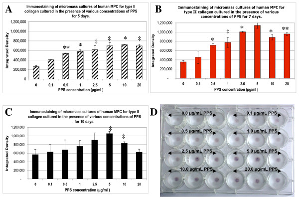
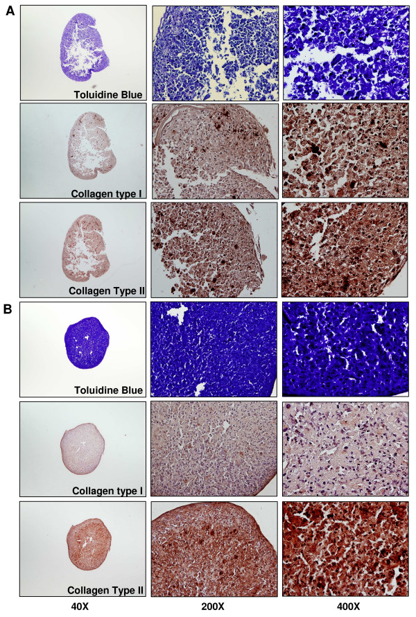
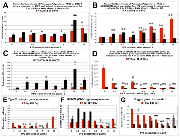
Human MPCs were maintained in monolayer, pellet or micromass cultures (MMC) for up to 10 days with PPS at concentrations of 0 to 20 microg/ml. MPC viability and proliferation was assessed using the WST-1 assay and 3H-thymidine incorporation into DNA, while apoptosis was monitored by flow cytometry. Proteoglycan (PG) biosynthesis was determined by 35SO42- incorporation and staining with Alcian blue. Proteoglycan and collagen type I and collagen type II deposition in pellet cultures was also examined by Toluidine blue and immunohistochemical staining, respectively. The production of hyaluronan (HA) by MPCs in MMC was assessed by ELISA. The relative outcome of PPS, HA, heparin or dextran sulfate (DS) on PG synthesis was compared in 5-day MMC. Gene expression of MPCs in 7-day and 10-day MMC was examined using real-time PCR. MPC differentiation was investigated by co-culturing with PPS in osteogenic or adipogenic inductive culture media for 28 days.
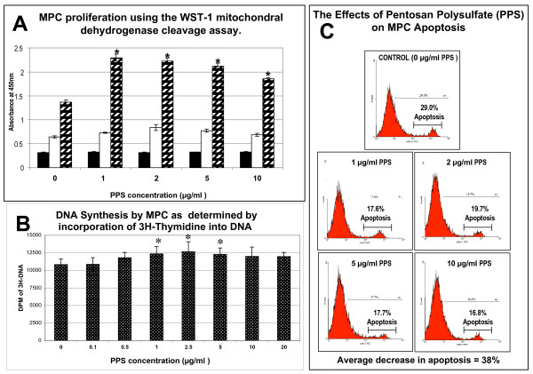
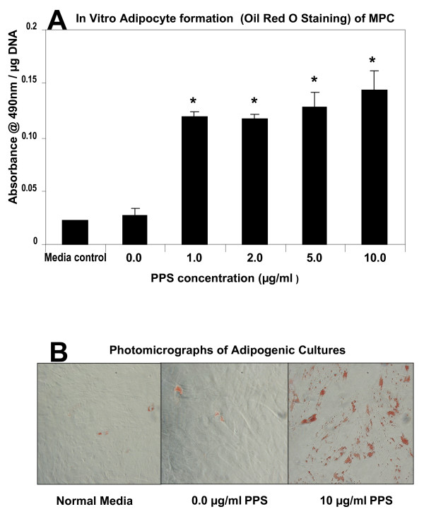
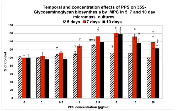
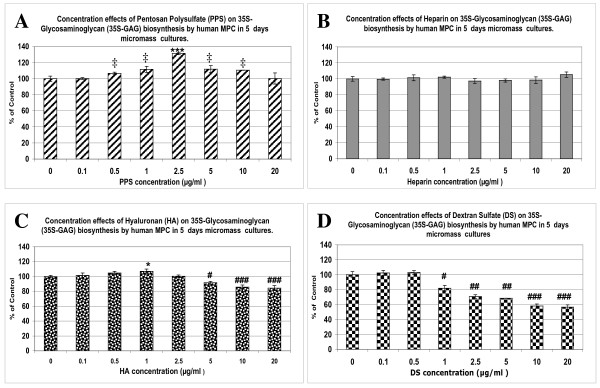
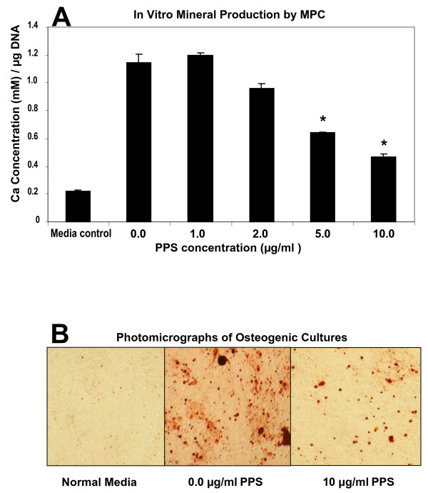
Significant MPC proliferation was evident by day 3 at PPS concentrations of 1 to 5 microg/ml (P < 0.01). In the presence of 1 to 10 microg/ml PPS, a 38% reduction in IL-4/IFNgamma-induced MPC apoptosis was observed. In 5-day MMC, 130% stimulation of PG synthesis occurred at 2.5 microg/ml PPS (P < 0.0001), while 5.0 microg/ml PPS achieved maximal stimulation in the 7-day and 10-day cultures (P < 0.05). HA and DS at > or = 5 microg/ml inhibited PG synthesis (P < 0.05) in 5-day MMC. Collagen type II deposition by MMC was significant at > or = 0.5 microg/ml PPS (P < 0.001 to 0.05). In MPC-PPS pellet cultures, more PG, collagen type II but less collagen type I was deposited than in controls. Real-time PCR results were consistent with the protein data. At 5 and 10 microg/ml PPS, MPC osteogenic differentiation was suppressed (P < 0.01).
This is the first study to demonstrate that PPS promotes MPC proliferation and chondrogenesis, offering new strategies for cartilage regeneration and repair in osteoarthritic joints.
本研究旨在确定抗骨关节炎药物戊聚糖多硫酸酯(PPS)是否影响间充质前体细胞(MPC)的增殖和分化。
用人 MPC 在单层、微球或微团培养物(MMC)中培养,浓度为 0 至 20μg/ml 的 PPS 存在 10 天。使用 WST-1 测定法和 3H-胸腺嘧啶掺入 DNA 来评估 MPC 的活力和增殖,而通过流式细胞术监测细胞凋亡。通过 35SO42-掺入和阿利新蓝染色来确定蛋白聚糖(PG)生物合成。通过甲苯胺蓝和免疫组织化学染色分别检查微球培养物中 PG 和胶原 I 型和胶原 II 型的沉积。通过 ELISA 评估 MPC 在 MMC 中透明质酸(HA)的产生。在 5 天 MMC 中比较 PPS、HA、肝素或葡聚糖硫酸盐(DS)对 PG 合成的相对作用。使用实时 PCR 检查 7 天和 10 天 MMC 中 MPC 的基因表达。通过与 PPS 共培养在成骨或成脂诱导培养基中培养 28 天来研究 MPC 分化。
在 1 至 5μg/ml 的 PPS 浓度下,MPC 增殖在第 3 天明显(P <0.01)。在 1 至 10μg/ml PPS 的存在下,观察到 IL-4/IFNgamma 诱导的 MPC 凋亡减少了 38%。在 5 天 MMC 中,2.5μg/ml PPS 导致 PG 合成增加了 130%(P <0.0001),而 5.0μg/ml PPS 在 7 天和 10 天培养物中达到最大刺激(P <0.05)。5 天 MMC 中 HA 和 DS 大于或等于 5μg/ml 抑制 PG 合成(P <0.05)。MPC 在大于或等于 0.5μg/ml PPS 时,在 MMC 中沉积的 II 型胶原增加(P <0.001 至 0.05)。在 MPC-PPS 微球培养物中,与对照相比,沉积的 PG、II 型胶原较多,但 I 型胶原较少。实时 PCR 结果与蛋白数据一致。在 5 和 10μg/ml PPS 下,MPC 成骨分化受到抑制(P <0.01)。
这是第一项证明 PPS 促进 MPC 增殖和软骨形成的研究,为骨关节炎关节中的软骨再生和修复提供了新的策略。