Center for Neuroproteomics and Biomarkers Research, Department of Psychiatry, McKnight Brain Institute of the University of Florida, Gainesville, FL 32610, USA.
BMC Neurosci. 2010 Feb 18;11:21. doi: 10.1186/1471-2202-11-21.
Autophagy, an intracellular response to stress, is characterized by double membrane cytosolic vesicles called autophagosomes. Prolonged autophagy is known to result in autophagic (Type II) cell death. This study examined the potential role of an autophagic response in cultured cerebellar granule neurons challenged with excitotoxin N-methyl-D-aspartate (NMDA).
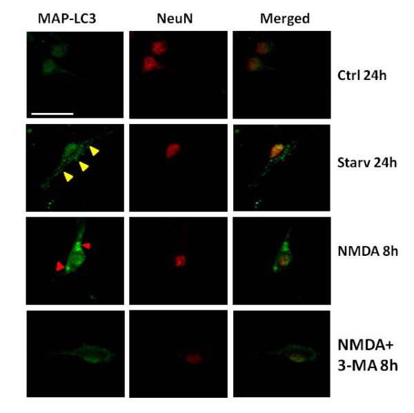
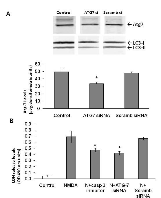
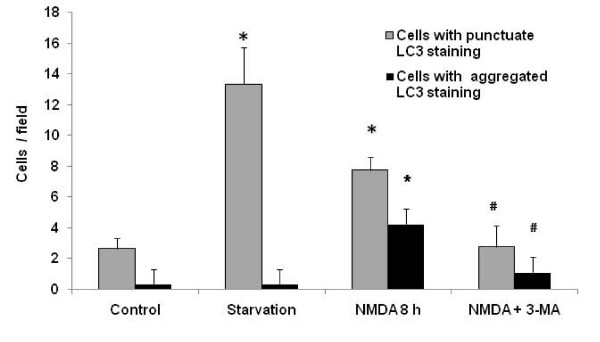
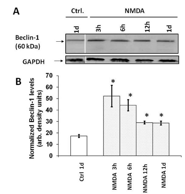
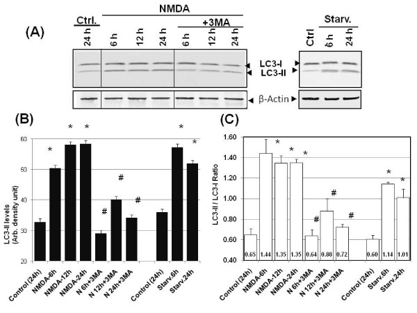
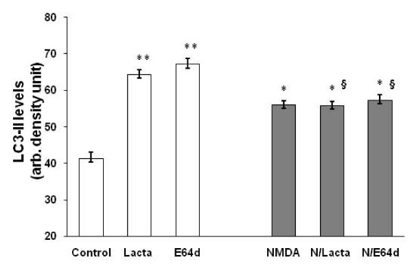
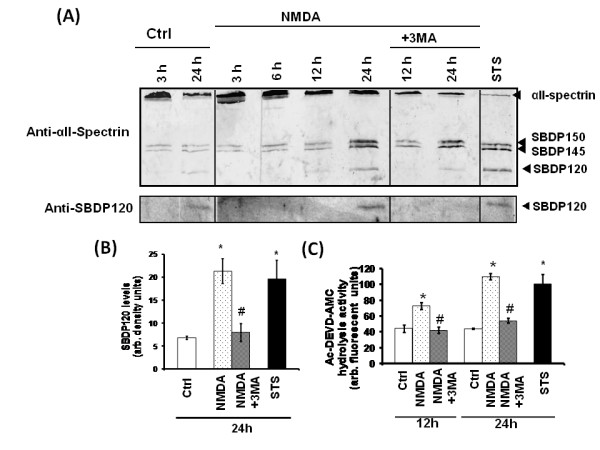
NMDA exposure induced light chain-3 (LC-3)-immunopositive and monodansylcadaverine (MDC) fluorescent dye-labeled autophagosome formation in both cell bodies and neurites as early as 3 hours post-treatment. Elevated levels of Beclin-1 and the autophagosome-targeting LC3-II were also observed following NMDA exposure. Prolonged exposure of the cultures to NMDA (8-24 h) generated MDC-, LC3-positive autophagosomal bodies, concomitant with the neurodegenerative phase of NMDA challenge. Lysosomal inhibition studies also suggest that NMDA-treatment diverted the autophagosome-associated LC3-II from the normal lysosomal degradation pathway. Autophagy inhibitor 3-methyladenine significantly reduced NMDA-induced LC3-II/LC3-I ratio increase, accumulation of autophagosomes, and suppressed NMDA-mediated neuronal death. ATG7 siRNA studies also showed neuroprotective effects following NMDA treatment.
Collectively, this study shows that autophagy machinery is robustly induced in cultured neurons subjected to prolonged exposure to excitotoxin, while autophagosome clearance by lysosomal pathway might be impaired. Our data further show that prolonged autophagy contributes to cell death in NMDA-mediated excitotoxicity.
自噬是一种对压力的细胞内反应,其特征是双层细胞质囊泡,称为自噬体。已知长期自噬会导致自噬(II 型)细胞死亡。本研究探讨了自噬反应在培养的小脑颗粒神经元受到兴奋性毒素 N-甲基-D-天冬氨酸(NMDA)挑战时的潜在作用。
NMDA 暴露后,在 3 小时的处理后,细胞体和突起中就出现了 LC-3 免疫阳性和单丹磺酰戊二醛(MDC)荧光染料标记的自噬体形成。NMDA 暴露后还观察到 Beclin-1 和自噬体靶向 LC3-II 的水平升高。培养物中 NMDA 的长时间暴露(8-24 小时)产生了 MDC、LC3 阳性自噬体小体,与 NMDA 挑战的神经退行性阶段同时发生。溶酶体抑制研究还表明,NMDA 处理使自噬体相关的 LC3-II 偏离了正常的溶酶体降解途径。自噬抑制剂 3-甲基腺嘌呤显著降低了 NMDA 诱导的 LC3-II/LC3-I 比值增加、自噬体积累,并抑制了 NMDA 介导的神经元死亡。ATG7 siRNA 研究也表明,NMDA 处理后具有神经保护作用。
总之,这项研究表明,在培养的神经元中,自噬机制在受到长时间暴露于兴奋性毒素时被强烈诱导,而溶酶体途径清除自噬体可能受损。我们的数据还表明,长期自噬会导致 NMDA 介导的兴奋性毒性中的细胞死亡。