Department of Molecular and Cellular Physiology, Stanford University, Stanford, California, USA.
Nat Struct Mol Biol. 2010 Mar;17(3):325-31. doi: 10.1038/nsmb.1764. Epub 2010 Feb 21.
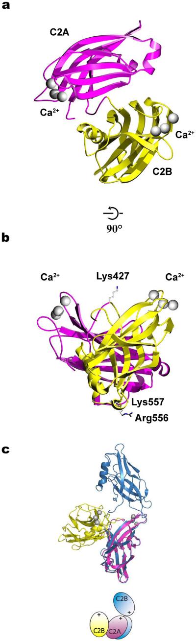
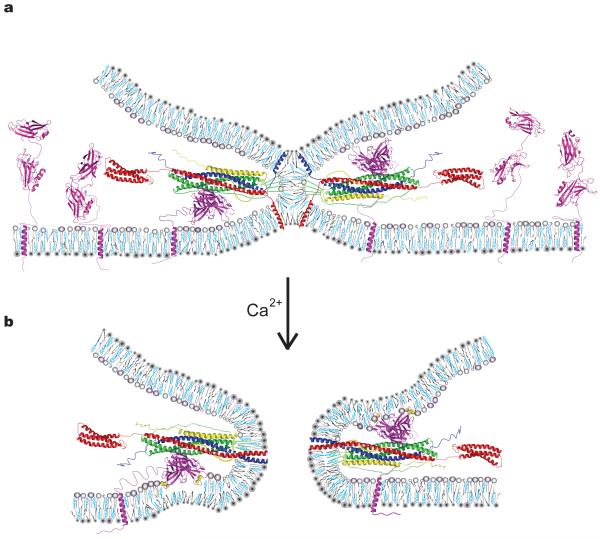
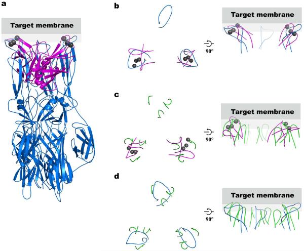
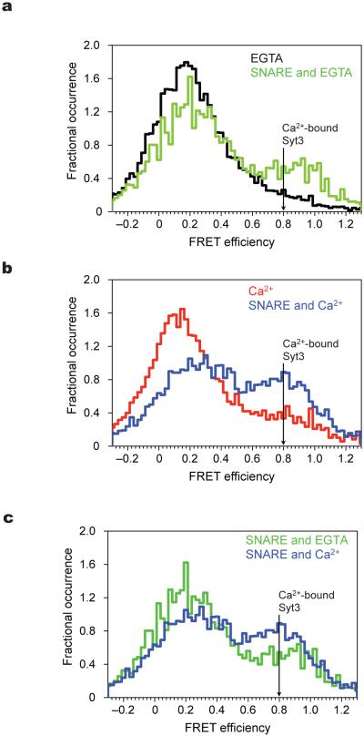
In neurons, SNAREs, synaptotagmin and other factors catalyze Ca(2+)-triggered fusion of vesicles with the plasma membrane. The molecular mechanism of this process, especially the interaction between synaptotagmin and SNAREs, remains an enigma. Here we characterized this interaction by single-molecule fluorescence microscopy and crystallography. The two rigid Ca(2+)-binding domains of synaptotagmin 3 (Syt3) undergo large relative motions in solution. Interaction with SNARE complex amplifies a particular state of the two domains that is further enhanced by Ca(2+). This state is represented by the first SNARE-induced Ca(2+)-bound crystal structure of a synaptotagmin fragment containing both domains. The arrangement of the Ca(2+)-binding loops of this structure of Syt3 matches that of SNARE-bound Syt1, suggesting a conserved feature of synaptotagmins. The loops resemble the membrane-interacting loops of certain viral fusion proteins in the postfusion state, suggesting unexpected similarities between both fusion systems.
在神经元中,SNARE 蛋白、突触融合蛋白和其他因子催化囊泡与质膜的 Ca2+触发融合。该过程的分子机制,尤其是突触融合蛋白与 SNARE 蛋白之间的相互作用,仍然是一个谜。在这里,我们通过单分子荧光显微镜和晶体学对这种相互作用进行了表征。突触融合蛋白 3(Syt3)的两个刚性 Ca2+结合结构域在溶液中会发生较大的相对运动。与 SNARE 复合物的相互作用放大了两个结构域的特定状态,而 Ca2+进一步增强了这种状态。这种状态由第一个 SNARE 诱导的 Ca2+结合的包含两个结构域的突触融合蛋白片段的晶体结构来表示。该 Syt3 结构的 Ca2+结合环的排列与 SNARE 结合的 Syt1 相匹配,表明突触融合蛋白具有保守特征。这些环类似于某些病毒融合蛋白在融合后状态下的膜相互作用环,表明这两种融合系统之间存在意想不到的相似性。