Department of Physics, North Carolina State University, Raleigh, North Carolina, USA.
Nat Struct Mol Biol. 2010 Mar;17(3):318-24. doi: 10.1038/nsmb.1763. Epub 2010 Feb 21.
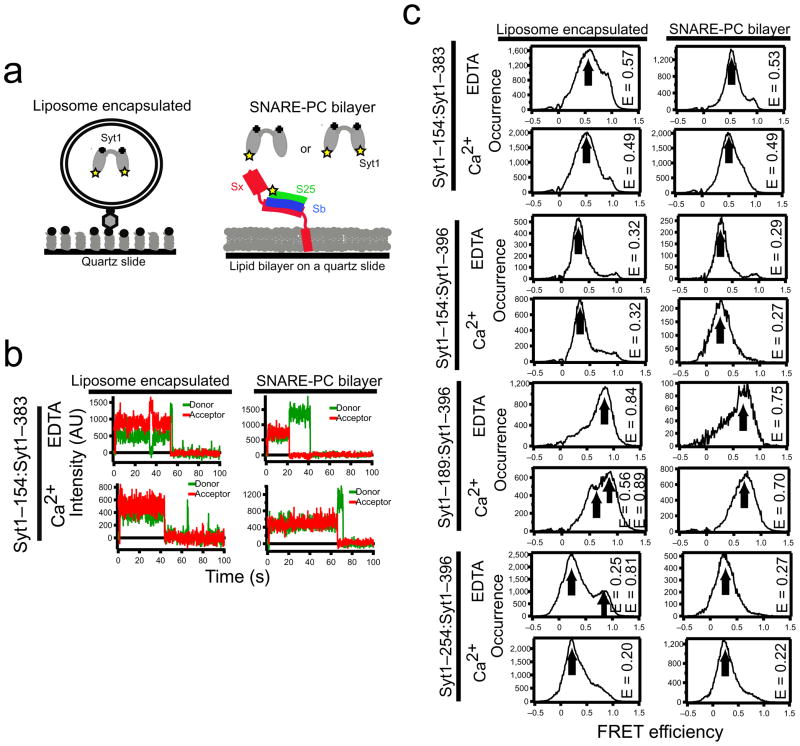
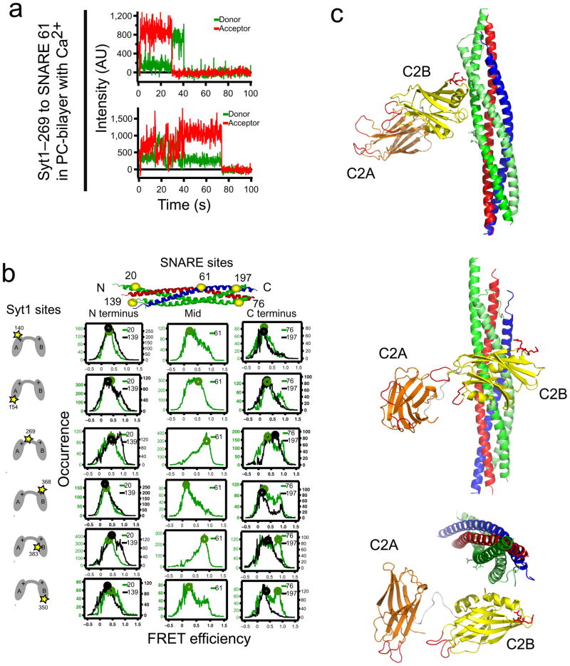
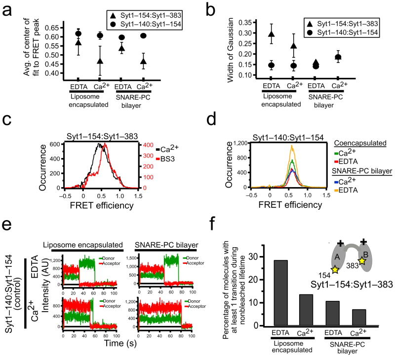
Synchronous neurotransmission is triggered when Ca(2+) binds to synaptotagmin 1 (Syt1), a synaptic-vesicle protein that interacts with SNAREs and membranes. We used single-molecule fluorescence resonance energy transfer (FRET) between synaptotagmin's two C2 domains to determine that their conformation consists of multiple states with occasional transitions, consistent with domains in random relative motion. SNARE binding results in narrower intrasynaptotagmin FRET distributions and less frequent transitions between states. We obtained an experimentally determined model of the elusive Syt1-SNARE complex using a multibody docking approach with 34 FRET-derived distances as restraints. The Ca(2+)-binding loops point away from the SNARE complex, so they may interact with the same membrane. The loop arrangement is similar to that of the crystal structure of SNARE-induced Ca(2+)-bound Syt3, suggesting a common mechanism by which the interaction between synaptotagmins and SNAREs aids in Ca(2+)-triggered fusion.
当钙离子与突触融合蛋白 1(Syt1)结合时,就会引发同步神经递质释放。Syt1 是一种突触囊泡蛋白,可与 SNARE 蛋白和膜相互作用。我们使用突触融合蛋白两个 C2 结构域之间的单分子荧光共振能量转移(FRET),确定其构象由多个状态组成,偶尔会发生状态间的转变,这与相对运动的域一致。SNARE 结合导致突触融合蛋白内 FRET 分布变窄,状态间的转变频率降低。我们使用多体对接方法,结合 34 个 FRET 衍生距离作为约束条件,获得了难以捉摸的 Syt1-SNARE 复合物的实验确定模型。钙结合环指向 SNARE 复合物的外侧,因此它们可能与同一膜相互作用。环的排列与 SNARE 诱导的 Ca2+结合 Syt3 的晶体结构相似,这表明 Syt 蛋白与 SNARE 蛋白之间的相互作用促进 Ca2+触发融合的机制可能是相似的。