Department of Molecular and Integrative Physiology, University of Illinois at Urbana-Champaign, Urbana, IL, USA.
Mol Neurodegener. 2010 Feb 9;5(1):9. doi: 10.1186/1750-1326-5-9.
Alpha-synuclein is a presynaptic protein with a proposed role in neurotransmission and dopamine homeostasis. Abnormal accumulation of alpha-synuclein aggregates in dopaminergic neurons of the substantia nigra is diagnostic of sporadic Parkinson's disease, and mutations in the protein are linked to early onset forms of the disease. The folded conformation of the protein varies depending upon its environment and other factors that are poorly understood. When bound to phospholipid membranes, alpha-synuclein adopts a helical conformation that mediates specific interactions with other proteins.
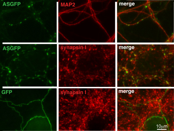
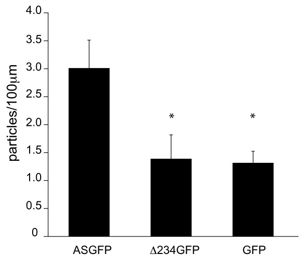
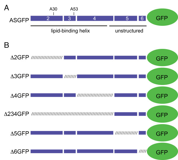
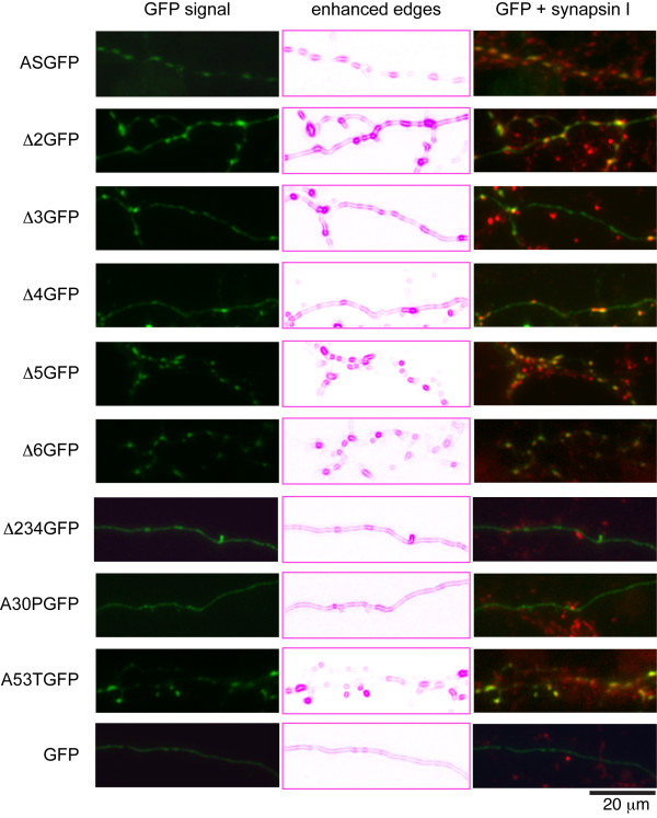
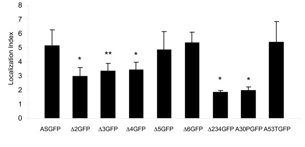
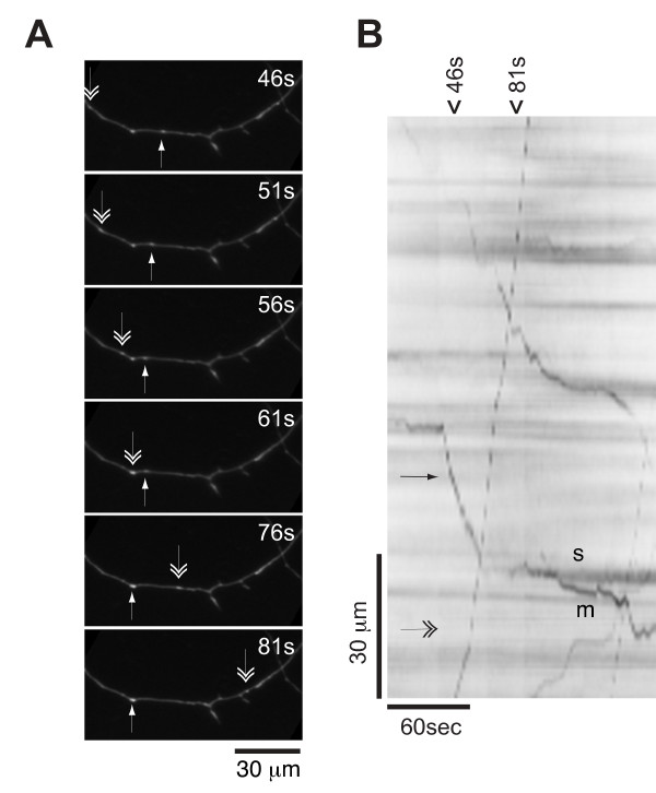
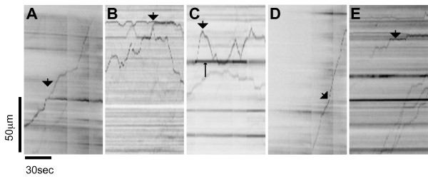
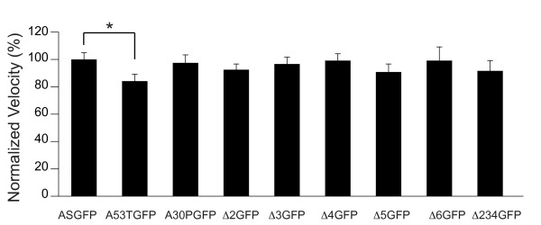
To investigate the role of the helical domain in transport and localization of alpha-synuclein, eGFP-tagged constructs were transfected into rat primary hippocampal neurons at 7 DIV. A series of constructs were analyzed in which each individual exon was deleted, for comparison to previous studies of lipid affinity and alpha-helix content. A53T and A30P substitutions, representing Parkinson's disease-associated variants, were analyzed as well. Single exon deletions within the lipid-binding N-terminal domain of alpha-synuclein (exons 2, 3, and 4) partially disrupted its presynaptic localization at 17-21 DIV, resulting in increased diffuse labeling of axons. Similar results were obtained for A30P, which exhibits decreased lipid binding, but not A53T. To examine whether differences in presynaptic enrichment were related to deficiencies in transport velocity, transport was visualized via live cell microscopy. Tagged alpha-synuclein migrated at a rate of 1.85 +/- 0.09 mum/s, consistent with previous reports, and single exon deletion mutants migrated at similar rates, as did A30P. Deletion of the entire N-terminal lipid-binding domain (Delta234GFP) did not significantly alter rates of particle movement, but decreased the number of moving particles. Only the A53TGFP mutant exhibited a significant decrease in transport velocity as compared to ASGFP.
These results support the hypothesis that presynaptic localization involves a mechanism that requires helical conformation and lipid binding. Conversely, the rate of axonal transport is not determined by lipid affinity and is not sufficient to account for differences in presynaptic localization of alpha-synuclein-eGFP variants.
α-突触核蛋白是一种突触前蛋白,据推测在神经递质传递和多巴胺稳态中发挥作用。在黑质多巴胺能神经元中异常聚集的α-突触核蛋白是散发性帕金森病的诊断标志,而该蛋白的突变与疾病的早发形式有关。该蛋白的折叠构象取决于其环境和其他因素,但这些因素知之甚少。当与磷脂膜结合时,α-突触核蛋白会采用螺旋构象,从而介导与其他蛋白的特异性相互作用。
为了研究螺旋结构域在α-突触核蛋白运输和定位中的作用,在 7 天分化(DIV)时将 eGFP 标记的构建体转染到大鼠原代海马神经元中。分析了一系列构建体,其中每个单独的外显子被删除,以与先前的脂质亲和力和α-螺旋含量研究进行比较。还分析了 A53T 和 A30P 取代,代表帕金森病相关变体。α-突触核蛋白的脂质结合 N 端结构域内的单个外显子缺失(外显子 2、3 和 4)部分破坏了其在 17-21 DIV 时的突触前定位,导致轴突中弥散标记增加。对于 A30P 也得到了类似的结果,A30P 表现出脂质结合减少,但不是 A53T。为了研究突触前富集的差异是否与运输速度的缺陷有关,通过活细胞显微镜观察运输。标记的α-突触核蛋白的迁移速度为 1.85 +/- 0.09 µm/s,与先前的报道一致,单个外显子缺失突变体以相似的速度迁移,A30P 也是如此。删除整个 N 端脂质结合结构域(Delta234GFP)并没有显著改变颗粒运动的速度,但减少了运动颗粒的数量。只有 A53TGFP 突变体与 ASGFP 相比,表现出明显的运输速度下降。
这些结果支持这样的假设,即突触前定位涉及一种需要螺旋构象和脂质结合的机制。相反,轴突运输的速度不是由脂质亲和力决定的,也不足以解释α-突触核蛋白-eGFP 变体在突触前定位的差异。