Imaging Science Laboratories, Translational and Molecular imaging Institute, Department of Radiology, Mount Sinai School of Med, New York, NY, USA.
J Cardiovasc Magn Reson. 2010 Mar 5;12(1):10. doi: 10.1186/1532-429X-12-10.
Atherosclerosis is a progressive disease that causes vascular remodeling that can be positive or negative. The evolution of arterial wall thickening and changes in lumen size under current "standard of care" in different arterial beds is unclear. The purpose of this study was to examine arterial remodeling and progression/regression of atherosclerosis in aorta and carotid arteries of individuals at risk for atherosclerosis normalized over a 1-year period.
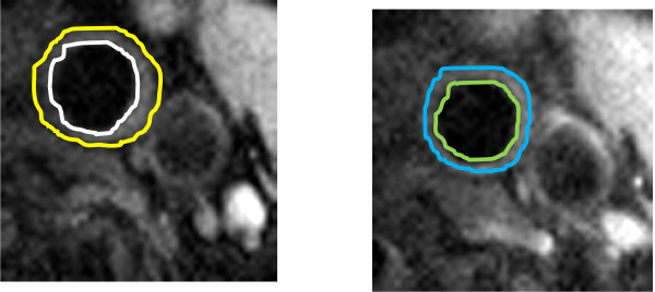
In this study, 28 patients underwent at least 2 black-blood in vivo cardiovascular magnetic resonance (CMR) scans of aorta and carotids over a one-year period (Mean 17.8 +/- 7.5 months). Clinical risk profiles for atherosclerosis and medications were documented and patients were followed by their referring physicians under current "standard of care" guidelines. Carotid and aortic wall lumen areas were matched across the time-points from cross-sectional images.
The wall area increased by 8.67%, 10.64%, and 13.24% per year (carotid artery, thoracic aorta and abdominal aorta respectively, p < 0.001). The lumen area of the abdominal aorta increased by 4.97% per year (p = 0.002), but the carotid artery and thoracic aorta lumen areas did not change significantly. The use of statin therapy did not change the rate of increase of wall area of carotid artery, thoracic and abdominal aorta, but decreased the rate of change of lumen area of carotid artery (-3.08 +/- 11.34 vs. 0.19 +/- 12.91 p < 0.05).
Results of this study of multiple vascular beds indicated that different vascular locations exhibited varying progression of atherosclerosis and remodeling as monitored by CMR.
动脉粥样硬化是一种进行性疾病,可导致血管重塑,其可呈阳性或阴性。在不同的动脉床中,目前“标准治疗”下动脉壁增厚和管腔大小变化的演变尚不清楚。本研究的目的是检查动脉粥样硬化风险个体的主动脉和颈动脉的动脉重塑和动脉粥样硬化的进展/消退,这些个体在一年内得到了正常化。
在这项研究中,28 名患者在一年内至少进行了 2 次主动脉和颈动脉的活体黑血心血管磁共振(CMR)扫描(平均 17.8 +/- 7.5 个月)。记录了动脉粥样硬化的临床风险概况和药物治疗,并根据当前的“标准治疗”指南由他们的主治医生对患者进行随访。通过横断面图像匹配颈动脉和主动脉壁管腔面积的时间点。
壁面积每年分别增加 8.67%、10.64%和 13.24%(颈动脉、胸主动脉和腹主动脉,p < 0.001)。腹主动脉的管腔面积每年增加 4.97%(p = 0.002),但颈动脉和胸主动脉管腔面积没有明显变化。他汀类药物治疗的使用并没有改变颈动脉、胸主动脉和腹主动脉壁面积增加的速度,但降低了颈动脉管腔面积变化的速度(-3.08 +/- 11.34 比 0.19 +/- 12.91,p < 0.05)。
这项多血管床研究的结果表明,不同的血管位置在 CMR 监测下表现出不同的动脉粥样硬化进展和重塑。