Schmitt-Gräff A, Krüger S, Bochard F, Gabbiani G, Denk H
Department of Pathology, University of Geneva, Switzerland.
Am J Pathol. 1991 May;138(5):1233-42.
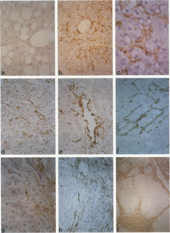
It has been suggested that perisinusoidal liver cells (PSC) play a pivotal role in the pathogenesis of fibrocontractive changes. Using light and electron microscopic immunolocalization techniques, a series of 207 normal and pathologic human liver specimens were evaluated for the expression of alpha smooth muscle (SM) actin and desmin in this and other nonparenchymal cell types. In normal adult liver tissue, PSCs were practically devoid of desmin and exceptionally stained for alpha-SM actin, whereas this actin isoform frequently was encountered in PSCs from the embryonic to the adolescent period. A broad spectrum of pathologic conditions was accompanied by the presence of alpha-SM actin containing PSCs; these were detected preferentially in periportal or perivenular zones according to the predominant location of the underlying hepatocellular damage. The occurrence of this PSC phenotype generally was associated with fibrogenesis and was in some cases detected earlier than overt collagen accumulation. Fibrous bands subdividing liver tissue in cirrhosis and focal nodular hyperplasia, as well as desmoplastic reaction to malignant tumors, contained alpha-SM actin-rich cells admixed with variable proportions of cells coexpressing desmin. In end stages, this population was less numerous than in active fibrotic or cirrhotic processes. Using immunogold electron microscopy, alpha-SM actin was localized in microfilament bundles of typical PSCs. Our results are compatible with the assumption that the appearance of alpha-SM actin and desmin-expressing myofibroblasts results at least in part from a phenotypic modulation of PSCs.
有人提出,肝血窦周细胞(PSC)在纤维收缩性改变的发病机制中起关键作用。利用光镜和电镜免疫定位技术,对207例正常和病理状态的人肝标本进行了评估,以检测α平滑肌(SM)肌动蛋白和结蛋白在这种及其他非实质细胞类型中的表达。在正常成人肝组织中,PSC几乎不表达结蛋白,α-SM肌动蛋白染色极淡,而在胚胎期至青春期的PSC中常可检测到这种肌动蛋白异构体。多种病理状态均伴有含α-SM肌动蛋白的PSC出现;根据潜在肝细胞损伤的主要部位,这些细胞优先在汇管区或中央静脉周围区域被检测到。这种PSC表型的出现通常与纤维生成有关,在某些情况下比明显的胶原沉积更早被检测到。在肝硬化和局灶性结节性增生中分隔肝组织的纤维带,以及对恶性肿瘤的促纤维增生反应,均含有富含α-SM肌动蛋白的细胞,并混有不同比例共表达结蛋白的细胞。在终末期,这一细胞群体比活跃的纤维化或肝硬化过程中的数量要少。利用免疫金电子显微镜技术,α-SM肌动蛋白定位于典型PSC的微丝束中。我们的结果与以下假设相符,即表达α-SM肌动蛋白和结蛋白的肌成纤维细胞的出现至少部分源于PSC的表型调节。