Schürch W, Skalli O, Seemayer T A, Gabbiani G
Am J Pathol. 1987 Jul;128(1):91-103.
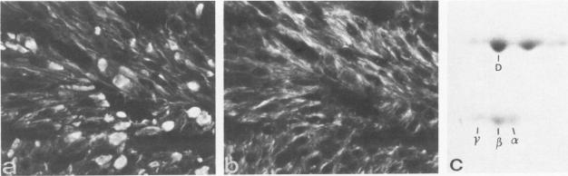
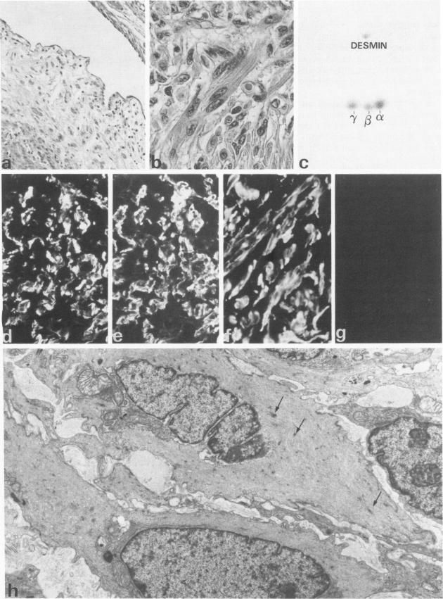
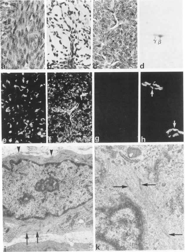
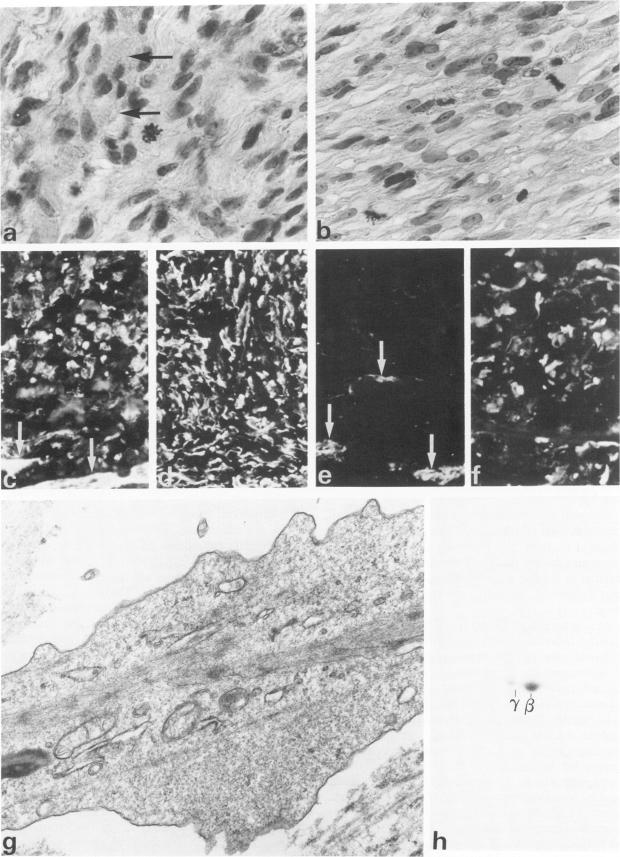
A series of 3 benign and 10 malignant smooth muscle (SM) neoplasms and of 2 malignant fibrous histiocytomas was examined by light microscopy, transmission electron microscopy, two-dimensional gel electrophoresis (2D-GE) and indirect immunofluorescence, using polyclonal monospecific or monoclonal antibodies to desmin, vimentin, cytokeratin, alpha-SM and alpha-sarcomeric (alpha-SR) actins. Benign neoplasms displayed typical light-microscopic features of SM, whereas leiomyosarcomas demonstrated variations in their histologic pattern. In 6 sarcomas, light microscopy suggested a SM differentiation, whereas in the other 4, a predominant nondistinctive spindle-cell pattern was observed. By transmission electron microscopy, all 13 neoplasms showed the minimal essential features of SM differentiation. Immunofluorescence disclosed heterogeneity of cytoskeletal protein expression: 5 neoplasms (3 benign and 2 malignant well-differentiated) expressed desmin, vimentin, and alpha-SM-actin; 2 malignant neoplasms expressed desmin and vimentin; 1 malignant neoplasm expressed desmin, vimentin and alpha-SR actin; 1 malignant neoplasm expressed vimentin and alpha-SR actin; and 4 malignant neoplasms expressed vimentin alone. By 2D-GE, 3 benign and 4 malignant SM neoplasms expressed alpha, beta, and gamma actins, and the remaining expressed only beta and gamma actins. The presence of alpha-SM actin in all benign neoplasms and in 2 well-differentiated leiomyosarcomas suggests that this actin isoform reflects a high degree of cellular differentiation. In 2 leiomyosarcomas, alpha-SR actin was detected by immunofluorescence, which suggested a skeletal muscle differentiation of these neoplasms. This study supports the assumption that leiomyosarcomas represent a heterogeneous group of neoplasms and furnishes new criteria for their characterization.
对3例良性和10例恶性平滑肌(SM)肿瘤以及2例恶性纤维组织细胞瘤进行了光镜、透射电镜、二维凝胶电泳(2D-GE)和间接免疫荧光检查,使用针对结蛋白、波形蛋白、细胞角蛋白、α-SM和α-肌节(α-SR)肌动蛋白的多克隆单特异性或单克隆抗体。良性肿瘤表现出平滑肌典型的光镜特征,而平滑肌肉瘤在组织学模式上存在差异。在6例肉瘤中,光镜提示平滑肌分化,而在另外4例中,观察到主要为无特征性的梭形细胞模式。通过透射电镜,所有13例肿瘤均显示出平滑肌分化的基本特征。免疫荧光揭示了细胞骨架蛋白表达的异质性:5例肿瘤(3例良性和2例恶性高分化)表达结蛋白、波形蛋白和α-SM肌动蛋白;2例恶性肿瘤表达结蛋白和波形蛋白;1例恶性肿瘤表达结蛋白、波形蛋白和α-SR肌动蛋白;1例恶性肿瘤表达波形蛋白和α-SR肌动蛋白;4例恶性肿瘤仅表达波形蛋白。通过2D-GE,3例良性和4例恶性平滑肌肿瘤表达α、β和γ肌动蛋白,其余仅表达β和γ肌动蛋白。所有良性肿瘤和2例高分化平滑肌肉瘤中α-SM肌动蛋白的存在表明这种肌动蛋白异构体反映了高度的细胞分化。在2例平滑肌肉瘤中,通过免疫荧光检测到α-SR肌动蛋白,提示这些肿瘤有骨骼肌分化。本研究支持平滑肌肉瘤代表一组异质性肿瘤的假设,并为其特征描述提供了新的标准。