CIRMM, Institute of Industrial Science, University of Tokyo, Komaba, Meguro-ku, Tokyo, Japan.
PLoS One. 2010 Mar 16;5(3):e9667. doi: 10.1371/journal.pone.0009667.
The ability to understand and locally control the morphogenesis of mammalian cells is a fundamental objective of cell and developmental biology as well as tissue engineering research. We present parylene-C (ParC) deposited on polydimethylsiloxane (PDMS) as a new substratum for in vitro advanced cell culture in the case of Human Hepatocarcinoma (HepG2) cells.
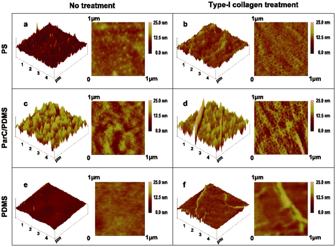
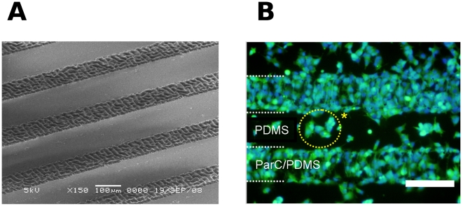
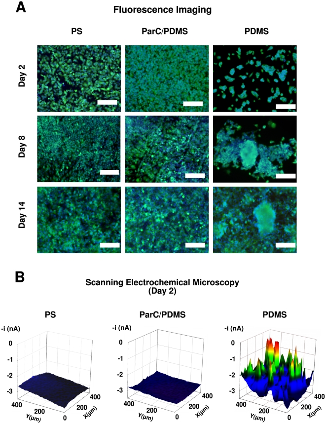
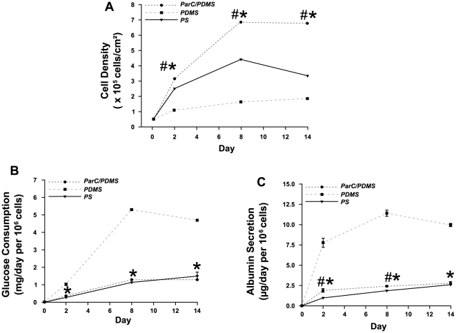
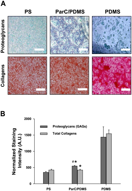
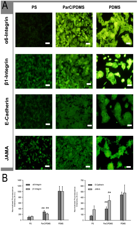
Our findings establish that the intrinsic properties of ParC-coated PDMS (ParC/PDMS) influence and modulate initial extracellular matrix (ECM; here, type-I collagen) surface architecture, as compared to non-coated PDMS substratum. Morphological changes induced by the presence of ParC on PDMS were shown to directly affect liver cell metabolic activity and the expression of transmembrane receptors implicated in cell adhesion and cell-cell interaction. These changes were characterized by atomic force microscopy (AFM), which elucidated differences in HepG2 cell adhesion, spreading, and reorganization into two- or three-dimensional structures by neosynthesis of ECM components. Local modulation of cell aggregation was successfully performed using ParC/PDMS micropatterns constructed by simple microfabrication.
CONCLUSION/SIGNIFICANCE: We demonstrated for the first time the modulation of HepG2 cells' behavior in relation to the intrinsic physical properties of PDMS and ParC, enabling the local modulation of cell spreading in a 2D or 3D manner by simple microfabrication techniques. This work will provide promising insights into the development of cell-based platforms that have many applications in the field of in vitro liver tissue engineering, pharmacology and therapeutics.
理解和局部控制哺乳动物细胞的形态发生是细胞和发育生物学以及组织工程研究的基本目标。我们提出了聚对二甲苯(ParC)沉积在聚二甲基硅氧烷(PDMS)上,作为人肝癌(HepG2)细胞体外高级细胞培养的新基质。
我们的研究结果表明,与未涂层的 PDMS 基质相比,ParC 涂层 PDMS(ParC/PDMS)的固有特性会影响和调节初始细胞外基质(ECM;这里是 I 型胶原蛋白)表面结构。PDMS 上存在 ParC 所诱导的形态变化被证明会直接影响肝细胞代谢活性和参与细胞黏附和细胞-细胞相互作用的跨膜受体的表达。原子力显微镜(AFM)对这些变化进行了表征,阐明了 HepG2 细胞黏附、铺展以及通过 ECM 成分的新合成重组成二维或三维结构的差异。通过简单的微制造技术,使用 ParC/PDMS 微图案成功地实现了局部细胞聚集的调制。
结论/意义:我们首次证明了 HepG2 细胞行为与 PDMS 和 ParC 的固有物理性质之间的关系的调制,通过简单的微制造技术以 2D 或 3D 方式实现细胞铺展的局部调制。这项工作将为基于细胞的平台的发展提供有前景的见解,这些平台在体外肝脏组织工程、药理学和治疗学等领域有许多应用。