Department of Psychiatry, Duke University Medical Center, Durham, North Carolina, USA.
Int Rev Psychiatry. 2009;21(4):394-409. doi: 10.1080/09540260902962198.
Cortical and subcortical hyperintensities in magnetic resonance imaging (MRI) scans are thought to represent areas of ischemic damage to brain tissue. Researchers have focused on the possible role these lesions may have in psychiatric disorders, including bipolar disorder. In 1997, the proposed 'vascular mania' diagnosis suggested utilizing not only the presence of strokes, but also confluent hyperintensities in its diagnostic criteria. This study was conducted to use meta-analytic techniques to investigate the association of hyperintensities and bipolar illness and to evaluate the current state of the literature.
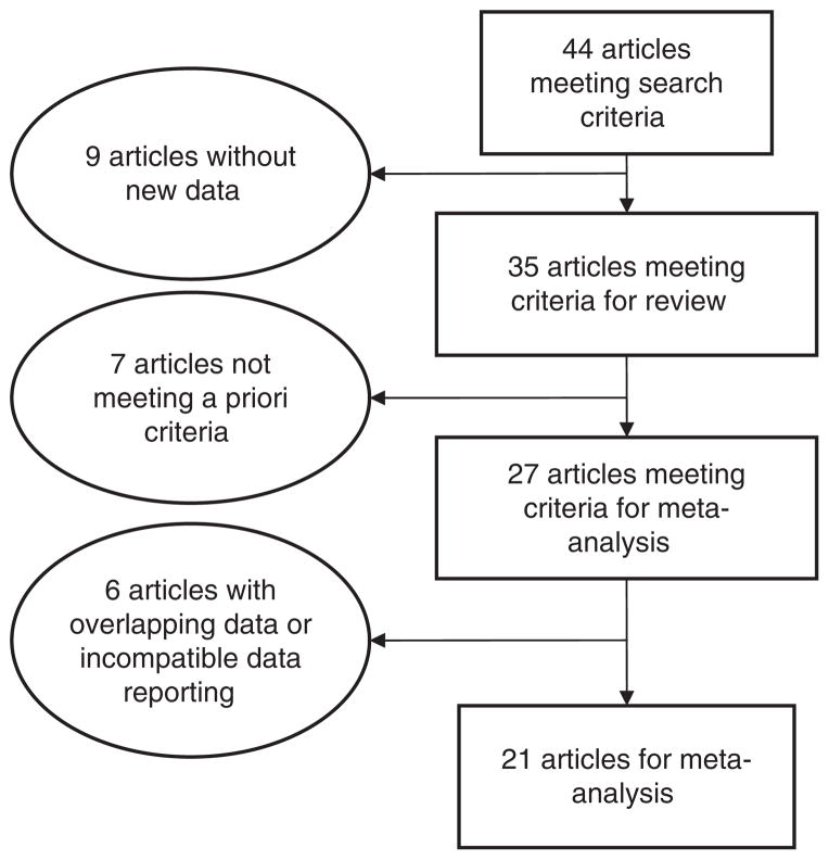
Using the PubMed and MEDLINE databases, we conducted a systematic literature search of studies investigating hyperintensities in subjects with bipolar disorder and controls or other psychiatric illnesses. We identified 44 publications from which 35 studies were included for review and 27 were selected for meta-analysis. Summary statistics of the prevalence were estimated through odds-ratios and confidence interval. Heterogeneity of the results across studies was tested using Q-statistics.
Meta-analysis identified an odds ratio of 2.5 (95% CI 1.9, 3.3) for hyperintensities in bipolar subjects compared to controls; however, there was significant heterogeneity among the studies (Q-statistics = 32; p = 0.04). This finding was most prominent for adolescents and children where the odds ratio was 5.7 (95% CI 2.3, 13.7). Deep white matter hyperintensities (odd ratio 3.2; 95% CI 2.2, 4.5) and subcortical grey matter hyperintensities (odds ratio 2.7; 95% CI 1.3, 2.9) were more strongly associated with bipolar subjects. There were no differences between bipolar subjects and controls for perivascular hyperintensities (odds ratio 1.3; 95% CI 0.8, 1.9). Though hyperintensities were numerically greater in bipolar subjects, meta-analysis did not demonstrate any significant differences between bipolar subjects and unipolar depression subjects (OR 1.6; 95% CI 0.9, 2.7) nor subjects with schizophrenia (OR 1.5; 95% CI 0.9, 2.7).
This meta-analysis continues to support the association of bipolar disorder and hyperintensities, especially in the deep white matter and subcortical grey matter. It also highlights the increased incidence in children and adolescence with bipolar disorder. However, hyperintensities are not specific to bipolar disorder, but appear at similar rates in unipolar depression and schizophrenia. Thus, the role of hyperintensities in the pathogenesis, pathophysiology, and treatment of bipolar disorder remains unclear. Further studies are required that are large enough to decrease the heterogeneity of the samples and MRI techniques, assess size and location of hyperintensities, and the impact on treatment response. Coordination with newer imaging techniques, such as diffusion tensor imaging (DTI) may be especially helpful in understanding the pathology of these lesions.
磁共振成像 (MRI) 扫描中的皮质和皮质下高信号被认为代表脑组织的缺血性损伤区域。研究人员专注于这些病变在包括双相情感障碍在内的精神疾病中的可能作用。1997 年,提出的“血管性躁狂”诊断建议不仅利用中风的存在,还利用其诊断标准中的融合性高信号。本研究旨在使用荟萃分析技术研究高信号与双相情感障碍之间的关联,并评估文献现状。
我们使用 PubMed 和 MEDLINE 数据库,对研究双相情感障碍患者和对照组或其他精神疾病患者高信号的文献进行了系统的文献检索。我们从 44 篇出版物中确定了 35 项研究进行综述,其中 27 项研究进行了荟萃分析。通过优势比和置信区间估计患病率的汇总统计数据。使用 Q 统计量测试研究之间的异质性。
荟萃分析确定,与对照组相比,双相情感障碍患者的高信号优势比为 2.5(95%置信区间 1.9, 3.3);然而,研究之间存在显著的异质性(Q 统计量=32;p=0.04)。这一发现在青少年和儿童中最为明显,优势比为 5.7(95%置信区间 2.3, 13.7)。深部白质高信号(优势比 3.2;95%置信区间 2.2, 4.5)和皮质下灰质高信号(优势比 2.7;95%置信区间 1.3, 2.9)与双相情感障碍患者的相关性更强。双相情感障碍患者和对照组之间的血管周围高信号无差异(优势比 1.3;95%置信区间 0.8, 1.9)。尽管双相情感障碍患者的高信号数值更大,但荟萃分析并未显示双相情感障碍患者与单相抑郁症患者(OR 1.6;95%置信区间 0.9, 2.7)或精神分裂症患者(OR 1.5;95%置信区间 0.9, 2.7)之间存在任何显著差异。
本荟萃分析继续支持双相情感障碍与高信号之间的关联,尤其是在深部白质和皮质下灰质中。它还突出了双相情感障碍在儿童和青少年中的发病率增加。然而,高信号并非双相情感障碍所特有,在单相抑郁症和精神分裂症中也以相似的比率出现。因此,高信号在双相情感障碍的发病机制、病理生理学和治疗中的作用仍不清楚。需要进一步的研究,这些研究的样本和 MRI 技术足够大,可以降低样本的异质性,评估高信号的大小和位置,以及对治疗反应的影响。与新的成像技术(如弥散张量成像 (DTI))的协调可能有助于理解这些病变的病理学。
