Department of Medicine, Imperial College London, London, UK.
PLoS One. 2010 May 24;5(5):e10777. doi: 10.1371/journal.pone.0010777.
Mycobacterium tuberculosis, the causative agent of tuberculosis, still represents a major public health threat in many countries. Bioluminescence, the production of light by luciferase-catalyzed reactions, is a versatile reporter technology with multiple applications both in vitro and in vivo. In vivo bioluminescence imaging (BLI) represents one of its most outstanding uses by allowing the non-invasive localization of luciferase-expressing cells within a live animal. Despite the extensive use of luminescent reporters in mycobacteria, the resultant luminescent strains have not been fully applied to BLI.
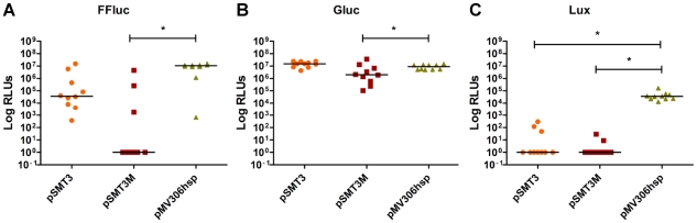
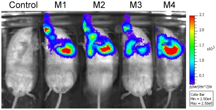
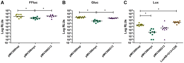
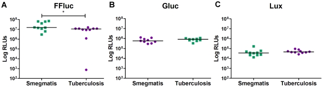
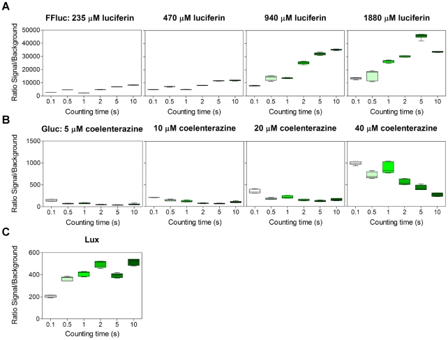
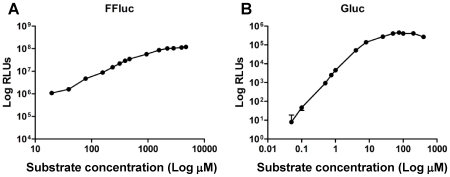
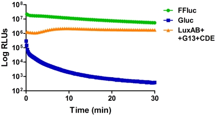
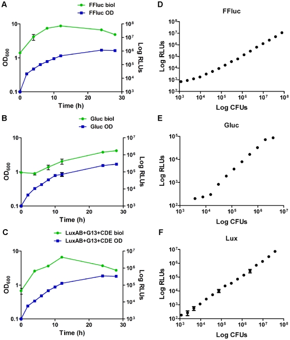
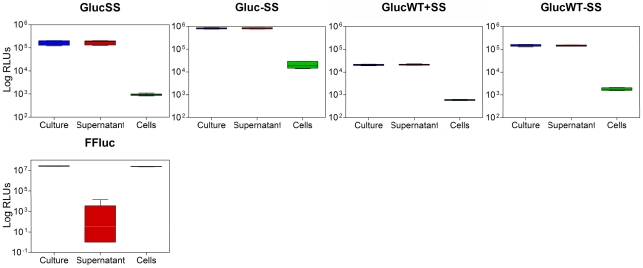
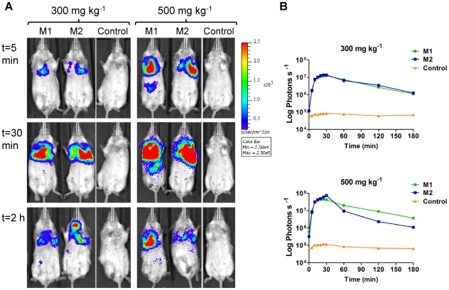
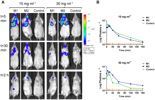
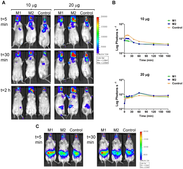
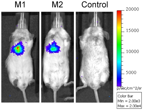
METHODOLOGY/PRINCIPAL FINDINGS: One of the main obstacles to the use of bioluminescence for in vivo imaging is the achievement of reporter protein expression levels high enough to obtain a signal that can be detected externally. Therefore, as a first step in the application of this technology to the study of mycobacterial infection in vivo, we have optimised the use of firefly, Gaussia and bacterial luciferases in mycobacteria using a combination of vectors, promoters, and codon-optimised genes. We report for the first time the functional expression of the whole bacterial lux operon in Mycobacterium tuberculosis and M. smegmatis thus allowing the development of auto-luminescent mycobacteria. We demonstrate that the Gaussia luciferase is secreted from bacterial cells and that this secretion does not require a signal sequence. Finally we prove that the signal produced by recombinant mycobacteria expressing either the firefly or bacterial luciferases can be non-invasively detected in the lungs of infected mice by bioluminescence imaging.
CONCLUSIONS/SIGNIFICANCE: While much work remains to be done, the finding that both firefly and bacterial luciferases can be detected non-invasively in live mice is an important first step to using these reporters to study the pathogenesis of M. tuberculosis and other mycobacterial species in vivo. Furthermore, the development of auto-luminescent mycobacteria has enormous ramifications for high throughput mycobacterial drug screening assays which are currently carried out either in a destructive manner using LuxAB or the firefly luciferase.
结核分枝杆菌是结核病的病原体,在许多国家仍然是一个主要的公共卫生威胁。生物发光是一种由荧光素酶催化反应产生光的多功能报告技术,在体外和体内都有多种应用。活体生物发光成像(BLI)是其最杰出的用途之一,它可以在活体动物体内非侵入性地定位表达荧光素酶的细胞。尽管在分枝杆菌中广泛使用发光报告基因,但这些发光菌株尚未完全应用于 BLI。
方法/主要发现:生物发光用于活体成像的主要障碍之一是实现足够高的报告蛋白表达水平,以获得可以外部检测到的信号。因此,作为将该技术应用于体内分枝杆菌感染研究的第一步,我们使用载体、启动子和密码子优化基因组合,优化了在分枝杆菌中使用萤火虫、Gaussia 和细菌荧光素酶的方法。我们首次报道了整个细菌 lux 操纵子在结核分枝杆菌和耻垢分枝杆菌中的功能表达,从而允许开发自发光分枝杆菌。我们证明 Gaussia 荧光素酶从细菌细胞中分泌出来,并且这种分泌不需要信号序列。最后,我们证明表达萤火虫或细菌荧光素酶的重组分枝杆菌产生的信号可以通过 BLI 在感染小鼠的肺部进行非侵入性检测。
结论/意义:虽然还有很多工作要做,但发现萤火虫和细菌荧光素酶都可以在活体小鼠中进行非侵入性检测,这是使用这些报告基因来研究结核分枝杆菌和其他分枝杆菌种在体内发病机制的重要第一步。此外,自发光分枝杆菌的开发对目前使用 LuxAB 或萤火虫荧光素酶以破坏性方式进行的高通量分枝杆菌药物筛选测定具有巨大影响。