Center for Molecular Imaging Research, Massachusetts General Hospital, Boston, MA, USA.
Eur Heart J. 2010 Aug;31(16):1975-84. doi: 10.1093/eurheartj/ehq237. Epub 2010 Jul 2.
Westernized countries face a growing burden of cardiovascular calcification and osteoporosis. Despite its vast clinical significance, the precise nature of this reciprocal relationship remains obscure. We hypothesize that cardiovascular calcification progresses with inflammation and inversely correlates with bone tissue mineral density (TMD).
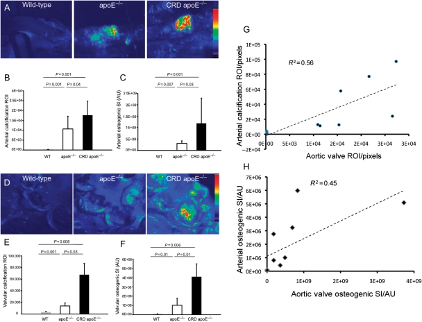
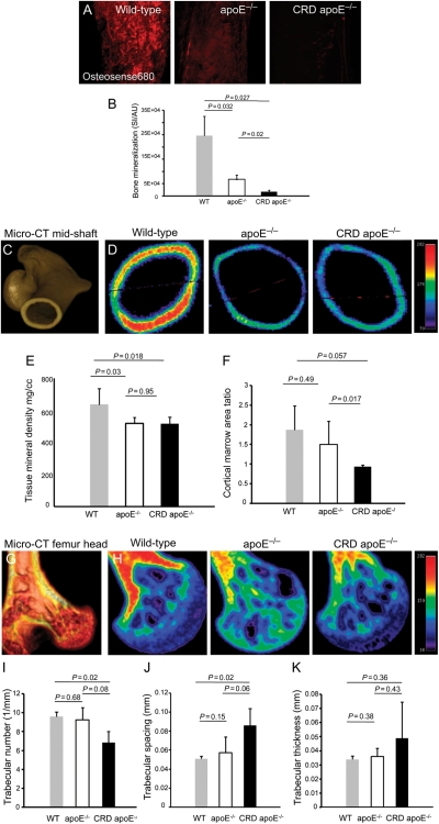
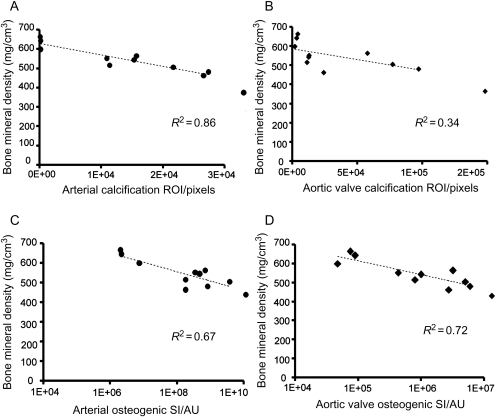
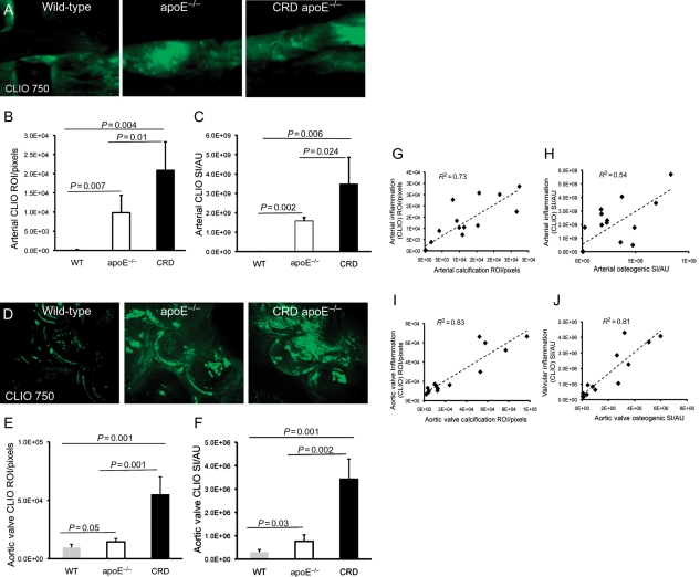
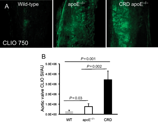
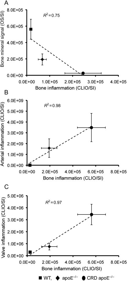
Arterial, valvular, and bone metabolism were visualized using near-infrared fluorescence (NIRF) molecular imaging agents, targeting macrophages and osteogenesis. We detected significant arterial and aortic valve calcification in apoE(-/-) mice with or without chronic renal disease (CRD, 30 weeks old; n = 28), correlating with the severity of atherosclerosis. We demonstrated decreases in osteogenic activity in the femurs of apoE(-/-) mice when compared with WT mice, which was further reduced with CRD. Three-dimensional micro-computed tomography imaging of the cortical and cancellous regions of femurs quantified structural remodelling and reductions in TMD in apoE(-/-) and CRD apoE(-/-) mice. We established significant correlations between arterial and valvular calcification and loss of TMD (R(2) = 0.67 and 0.71, respectively). Finally, we performed macrophage-targeted molecular imaging to explore a link between inflammation and osteoporosis in vivo. Although macrophage burden, visualized as uptake of NIRF-conjugated iron nanoparticles, was directly related to the degree of arterial and valvular inflammation and calcification, the same method inversely correlated inflammation with TMD (R(2) = 0.73; 0.83; 0.75, respectively).
This study provides direct in vivo evidence that in arteries and aortic valves, macrophage burden and calcification associate with each other, whereas inflammation inversely correlates with bone mineralization. Thus, understanding inflammatory signalling mechanisms may offer insight into selective abrogation of divergent calcific phenomena.
西化国家面临着心血管钙化和骨质疏松症负担日益加重的问题。尽管其具有广泛的临床意义,但这种相互关系的确切性质仍不清楚。我们假设心血管钙化是随着炎症进展的,并与骨组织矿物质密度(TMD)呈负相关。
使用近红外荧光(NIRF)分子成像剂对动脉、瓣膜和骨代谢进行可视化,靶向巨噬细胞和成骨细胞。我们在有或没有慢性肾脏病(CRD,30 周龄;n=28)的 apoE(-/-)小鼠中检测到明显的动脉和主动脉瓣钙化,与动脉粥样硬化的严重程度相关。我们发现与 WT 小鼠相比,apoE(-/-)小鼠的股骨成骨活性降低,而 CRD 进一步降低。股骨皮质和松质区域的三维微计算机断层扫描成像定量了 apoE(-/-)和 CRD apoE(-/-)小鼠的结构重塑和 TMD 降低。我们建立了动脉和瓣膜钙化与 TMD 丧失之间的显著相关性(R(2)分别为 0.67 和 0.71)。最后,我们进行了巨噬细胞靶向的分子成像,以探索体内炎症与骨质疏松症之间的联系。尽管动脉和瓣膜炎症和钙化程度与巨噬细胞负荷(表现为 NIRF 缀合铁纳米颗粒的摄取)直接相关,但相同的方法将炎症与 TMD 呈负相关(R(2)分别为 0.73;0.83;0.75)。
这项研究提供了直接的体内证据,表明在动脉和主动脉瓣中,巨噬细胞负荷和钙化相互关联,而炎症与骨矿化呈负相关。因此,了解炎症信号机制可能为选择性阻断不同的钙化现象提供启示。