Centre for Women's Health Research, Monash Institute of Medical Research and Monash University Department of Obstetrics and Gynaecology, Monash Medical Centre, 246 Clayton Rd, Clayton, VIC 3168, Australia.
Reprod Biol Endocrinol. 2010 Jul 8;8:84. doi: 10.1186/1477-7827-8-84.
It has been hypothesised that increased VEGF-D expression may be an independent prognostic factor for endometrial cancer progression and lymph node metastasis; however, the mechanism by which VEGF-D may promote disease progression in women with endometrial cancer has not been investigated. Our aim was to describe the distribution of lymphatic vessels in mouse uterus and to examine the effect of VEGF-D over-expression on these vessels in a model of endometrial cancer. We hypothesised that VEGF-D over-expression would stimulate growth of new lymphatic vessels into the endometrium, thereby contributing to cancer progression.
We initially described the distribution of lymphatic vessels (Lyve-1, podoplanin, VEGFR-3) and VEGF-D expression in the mouse uterus during the estrous cycle, early pregnancy and in response to estradiol-17beta and progesterone using immunohistochemistry. We also examined the effects of VEGF-D over-expression on uterine vasculature by inoculating uterine horns in NOD SCID mice with control or VEGF-D-expressing 293EBNA tumor cells.
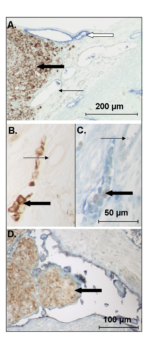
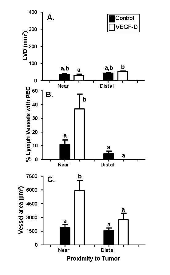
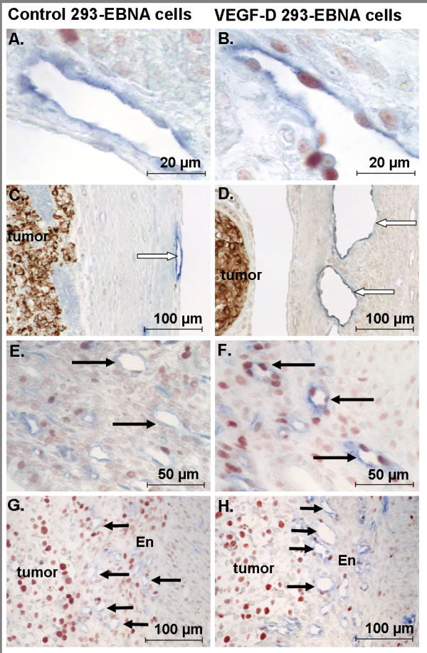
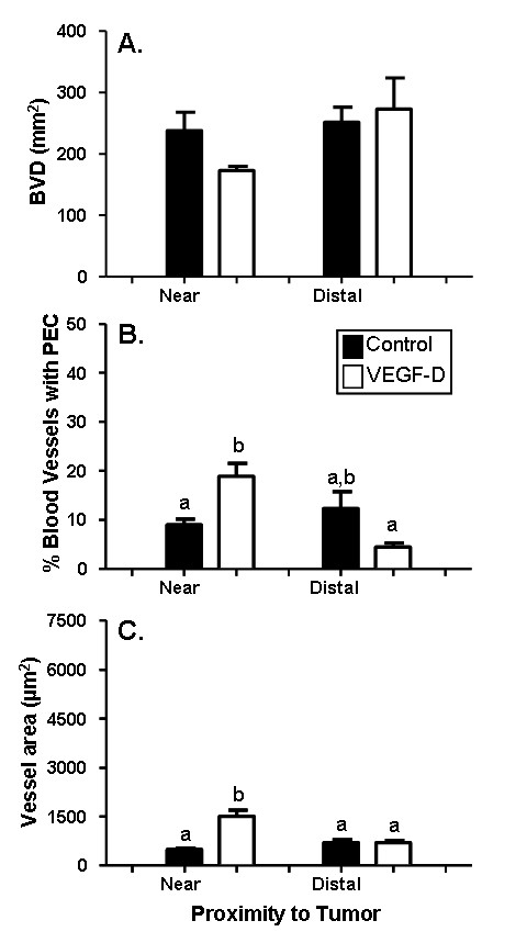
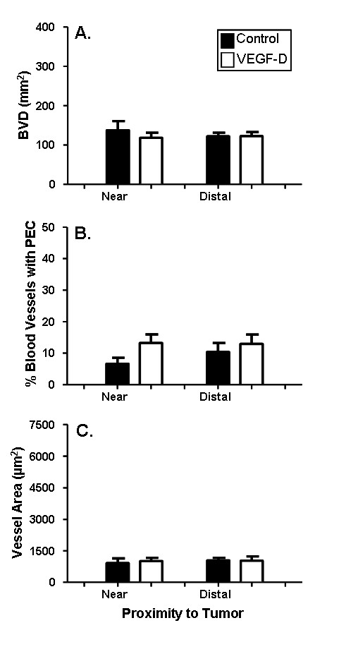
Lymphatic vessels positive for the lymphatic endothelial cell markers Lyve-1, podoplanin and VEGFR-3 profiles were largely restricted to the connective tissue between the myometrial circular and longitudinal muscle layers; very few lymphatic vessel profiles were observed in the endometrium. VEGF-D immunostaining was present in all uterine compartments (epithelium, stroma, myometrium), although expression was generally low. VEGF-D immunoexpression was slightly but significantly higher in estrus relative to diestrus; and in estradiol-17beta treated mice relative to vehicle or progesterone treated mice. The presence of VEGF-D over-expressing tumor cells did not induce endometrial lymphangiogenesis, although changes were observed in existing vessel profiles. For myometrial lymphatic and endometrial blood vessels, the percentage of profiles containing proliferating endothelial cells, and the cross sectional area of vessel profiles were significantly increased in response to VEGF-D in comparison to control tumor cells. In contrast, no significant changes were noted in myometrial blood vessels. In addition, examples of invading cells or tumor emboli were observed in mice receiving VEGF-D expressing 293EBNA cells.
These results illustrate that VEGF-D over-expression has differential effects on the uterine vasculature. These effects may facilitate VEGF-D's ability to promote endometrial cancer metastasis and disease progression.
已有假说认为,VEGF-D 表达增加可能是子宫内膜癌进展和淋巴结转移的独立预后因素;然而,VEGF-D 促进子宫内膜癌患者疾病进展的机制尚未得到研究。我们的目的是描述小鼠子宫中淋巴管的分布,并在子宫内膜癌模型中研究 VEGF-D 过表达对这些血管的影响。我们假设 VEGF-D 过表达会刺激新的淋巴管生长到子宫内膜中,从而促进癌症的进展。
我们最初使用免疫组织化学方法描述了在动情周期、早期妊娠以及雌二醇-17β和孕激素作用下,小鼠子宫中淋巴管(Lyve-1、podoplanin、VEGFR-3)的分布和 VEGF-D 的表达。我们还通过将对照或 VEGF-D 表达的 293EBNA 肿瘤细胞接种到 NOD SCID 小鼠的子宫角,研究了 VEGF-D 过表达对子宫血管的影响。
Lyve-1、podoplanin 和 VEGFR-3 等淋巴管内皮细胞标志物阳性的淋巴管主要局限于子宫环形和纵向平滑肌层之间的结缔组织中;子宫内膜中很少观察到淋巴管形态。VEGF-D 免疫染色存在于所有子宫腔(上皮、基质、子宫肌层),尽管表达通常较低。与间情期相比,动情期的 VEGF-D 免疫表达略高,但与雌二醇-17β处理的小鼠相比,与载体或孕激素处理的小鼠相比,VEGF-D 免疫表达略高。存在过表达 VEGF-D 的肿瘤细胞并不会诱导子宫内膜淋巴管生成,尽管观察到现有血管形态发生了变化。与对照肿瘤细胞相比,VEGF-D 可显著增加肌层淋巴管和子宫内膜血管中增殖内皮细胞的形态比例和血管形态的横截面积。相比之下,子宫血管没有明显变化。此外,在接受表达 VEGF-D 的 293EBNA 细胞的小鼠中观察到了浸润细胞或肿瘤栓子的例子。
这些结果表明,VEGF-D 过表达对子宫血管有不同的影响。这些影响可能有助于 VEGF-D 促进子宫内膜癌转移和疾病进展的能力。