• 文献检索
  • 文档翻译
  • 深度研究
  • 学术资讯
  • Suppr Zotero 插件Zotero 插件
  • 邀请有礼
  • 套餐&价格
  • 历史记录
应用&插件
Suppr Zotero 插件Zotero 插件浏览器插件Mac 客户端Windows 客户端微信小程序
定价
高级版会员购买积分包购买API积分包
服务
文献检索文档翻译深度研究API 文档MCP 服务
关于我们
关于 Suppr公司介绍联系我们用户协议隐私条款
关注我们

Suppr 超能文献

核心技术专利:CN118964589B侵权必究
粤ICP备2023148730 号-1Suppr @ 2026

文献检索

告别复杂PubMed语法,用中文像聊天一样搜索,搜遍4000万医学文献。AI智能推荐,让科研检索更轻松。

立即免费搜索

文件翻译

保留排版,准确专业,支持PDF/Word/PPT等文件格式,支持 12+语言互译。

免费翻译文档

深度研究

AI帮你快速写综述,25分钟生成高质量综述,智能提取关键信息,辅助科研写作。

立即免费体验

采用两种激光波长的超广角扫描激光检眼镜(Optomap)成像的脉络膜色素性病变

Choroidal pigmented lesions imaged by ultra-wide-field scanning laser ophthalmoscopy with two laser wavelengths (Optomap).

作者信息

Kernt Marcus, Schaller Ulrich C, Stumpf Carmen, Ulbig Michael W, Kampik Anselm, Neubauer Aljoscha S

机构信息

Department of Ophthalmology, Ludwig-Maximilians-University, Munich, Germany.

出版信息

Clin Ophthalmol. 2010 Jul 30;4:829-36. doi: 10.2147/opth.s11864.

DOI:10.2147/opth.s11864
PMID:20689737
原文链接:https://pmc.ncbi.nlm.nih.gov/articles/PMC2915871/
Abstract

PURPOSE

Clinical differentiation of choroidal pigmented lesions is sometimes difficult. Choroidal melanoma is the most prevalent primary neoplasia among malignant ocular tumors, and metastasis often occurs before the primary tumor is diagnosed. Therefore, early detection is essential. We investigated the imaging properties of clinically diagnosed melanocytic choroidal tumors using a nonmydriatic ultra-wide-field scanning laser ophthalmoscope (SLO) with two laser wavelengths to distinguish benign from malignant lesions. Repeated standardized ultrasound (US) evaluation provided reference standard.

METHODS

In a consecutive series of 49 patients with clinically diagnosed melanocytic choroidal tumors in one eye, 29 had established melanoma (defined by proven growth on repeated US follow-up) and 20 had nevi (defined by no malignancy according to clinical, US, and growth characteristics for at least 2 years). All patients underwent clinical examination, undilated Optomap((R)) (Optos PLC, Dunfermline, Fife, Scotland, UK) imaging, standardized US examination, and standard retinal photography. Measurements of the tumor base using the Optomap software were compared with US B-scan measurements. Imaging characteristics from the SLO images were correlated with the structural findings in the two patient groups.

RESULTS

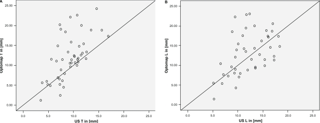
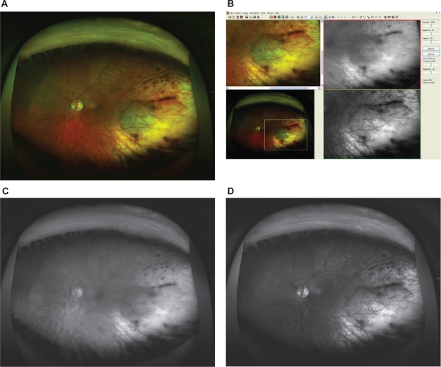
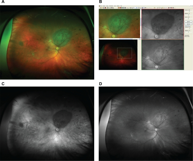
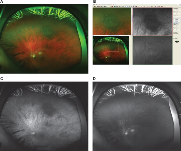
Measurements of tumor base correlated well between SLO and US with r = 0.61 (T-direction) and r = 0.51 (L-direction). On SLO imaging, typical malignant lesions appeared dark on the red laser channel and bright on the green laser channel. Based on those simple binary characteristics, a sensitivity of 76% at a specificity of 70% was obtained for a correct classification of lesions. When analogous to clinical examination lesion size, margin touching the optic disc, and existence of subretinal fluid were additionally considered, 90% sensitivity at 82% specificity was obtained.

CONCLUSIONS

In this first, limited series, nonmydriatic SLO imaging with two laser wavelengths permitted to differentiate malignant ocular tumors from nonmalignant lesions with high diagnostic accuracy. Additional parameters may further enhance diagnostic properties, but larger patient series are required to validate our findings and prove the diagnostic properties.

摘要

目的

脉络膜色素性病变的临床鉴别有时存在困难。脉络膜黑色素瘤是恶性眼肿瘤中最常见的原发性肿瘤,且转移常发生在原发性肿瘤被诊断之前。因此,早期检测至关重要。我们使用具有两种激光波长的免散瞳超广角扫描激光检眼镜(SLO)研究了临床诊断的脉络膜黑色素细胞肿瘤的成像特性,以区分良性和恶性病变。重复的标准化超声(US)评估提供了参考标准。

方法

在连续的49例单眼临床诊断为脉络膜黑色素细胞肿瘤的患者系列中,29例已确诊为黑色素瘤(根据重复的超声随访证实有生长来定义),20例为痣(根据临床、超声和生长特征至少2年无恶性来定义)。所有患者均接受了临床检查、未散瞳的Optomap(R)(Optos PLC,邓弗姆林,法夫,苏格兰,英国)成像、标准化超声检查和标准视网膜摄影。使用Optomap软件测量肿瘤基底并与超声B超测量结果进行比较。SLO图像的成像特征与两组患者的结构发现相关。

结果

SLO和US之间肿瘤基底测量的相关性良好,T方向r = 0.61,L方向r = 0.51。在SLO成像上,典型的恶性病变在红色激光通道上呈暗色,在绿色激光通道上呈亮色。基于这些简单的二元特征,病变正确分类的敏感性为76%,特异性为70%。当另外考虑类似于临床检查的病变大小、边缘接触视盘以及视网膜下液的存在时,敏感性为90%,特异性为82%。

结论

在这个首个有限的系列研究中,具有两种激光波长的免散瞳SLO成像能够以高诊断准确性区分恶性眼肿瘤和非恶性病变。其他参数可能会进一步提高诊断性能,但需要更大的患者系列来验证我们的发现并证明诊断性能。

https://cdn.ncbi.nlm.nih.gov/pmc/blobs/8229/2915871/3fbdf9295229/opth-4-829f4.jpg
https://cdn.ncbi.nlm.nih.gov/pmc/blobs/8229/2915871/42b48ffe450a/opth-4-829f1.jpg
https://cdn.ncbi.nlm.nih.gov/pmc/blobs/8229/2915871/6dd51eb0e651/opth-4-829f2.jpg
https://cdn.ncbi.nlm.nih.gov/pmc/blobs/8229/2915871/9b6bd257689c/opth-4-829f3.jpg
https://cdn.ncbi.nlm.nih.gov/pmc/blobs/8229/2915871/3fbdf9295229/opth-4-829f4.jpg

相似文献

1
Choroidal pigmented lesions imaged by ultra-wide-field scanning laser ophthalmoscopy with two laser wavelengths (Optomap).采用两种激光波长的超广角扫描激光检眼镜(Optomap)成像的脉络膜色素性病变
Clin Ophthalmol. 2010 Jul 30;4:829-36. doi: 10.2147/opth.s11864.
2
Role of wide-field autofluorescence imaging and scanning laser ophthalmoscopy in differentiation of choroidal pigmented lesions.广角自发荧光成像和扫描激光检眼镜在脉络膜色素性病变鉴别诊断中的作用。
Int J Ophthalmol. 2014 Aug 18;7(4):697-703. doi: 10.3980/j.issn.2222-3959.2014.04.21. eCollection 2014.
3
Assessment of diabetic retinopathy using nonmydriatic ultra-widefield scanning laser ophthalmoscopy (Optomap) compared with ETDRS 7-field stereo photography.应用非散瞳超广角激光扫描检眼镜(Optomap)评估糖尿病性视网膜病变与 ETDRS 7 区域立体摄影比较。
Diabetes Care. 2012 Dec;35(12):2459-63. doi: 10.2337/dc12-0346. Epub 2012 Aug 21.
4
Nonmydriatic ultra-wide-field scanning laser ophthalmoscopy (Optomap) versus two-field fundus photography in diabetic retinopathy.非散瞳超广角激光扫描检眼镜(Optomap)与两种眼底照相在糖尿病视网膜病变中的比较。
Ophthalmologica. 2014;231(1):31-6. doi: 10.1159/000355092. Epub 2013 Nov 13.
5
Ultra-wide-field imaging of choroidal melanoma before and after proton beam radiation therapy.脉络膜黑色素瘤质子束放射治疗前后的超广角成像。
Eur J Ophthalmol. 2020 Nov;30(6):1397-1402. doi: 10.1177/1120672119873210. Epub 2019 Aug 30.
6
Characterization of Choroidal Nevi with Dark-Field Infrared Scanning Laser Ophthalmoscopy.暗场红外扫描激光检眼镜对脉络膜痣的特征分析
Ophthalmol Retina. 2019 Aug;3(8):703-708. doi: 10.1016/j.oret.2019.03.011. Epub 2019 Mar 21.
7
Comparative study between fundus autofluorescence and red reflectance imaging of choroidal nevi using ultra-wide-field scanning laser ophthalmoscopy.使用超广角扫描激光检眼镜对脉络膜痣进行眼底自发荧光与红色反射成像的对比研究。
Retina. 2015 Jun;35(6):1202-10. doi: 10.1097/IAE.0000000000000463.
8
Nonmydriatic screening for diabetic retinopathy by ultra-widefield scanning laser ophthalmoscopy (Optomap).通过超广角扫描激光检眼镜(Optomap)进行糖尿病视网膜病变的免散瞳筛查。
Graefes Arch Clin Exp Ophthalmol. 2008 Feb;246(2):229-35. doi: 10.1007/s00417-007-0631-4. Epub 2007 Jul 11.
9
[Diabetic retinopathy: comparison of the diagnostic features of ultra-widefield scanning laser ophthalmoscopy Optomap with ETDRS 7-field fundus photography].[糖尿病视网膜病变:超广角扫描激光检眼镜Optomap与ETDRS七视野眼底摄影诊断特征的比较]
Ophthalmologe. 2011 Feb;108(2):117-23. doi: 10.1007/s00347-010-2226-4.
10
Screening for common eye diseases in the elderly with Optos ultra-wide-field scanning laser ophthalmoscopy: a pilot study with focus on ocular toxoplasmosis.应用 Optos 超广角激光扫描检眼镜筛查老年人常见眼病:以眼弓形虫病为重点的初步研究。
Int Ophthalmol. 2021 May;41(5):1573-1584. doi: 10.1007/s10792-020-01683-z. Epub 2021 Mar 16.

引用本文的文献

1
Measurement of Melanocytic Choroidal Lesions: Ultrasound Versus Ultrawide-Field Fundus Imaging System.脉络膜黑素细胞性病变的测量:超声与超广角眼底成像系统对比
Cancers (Basel). 2025 Feb 14;17(4):642. doi: 10.3390/cancers17040642.
2
Pixel Intensity to Estimate Choroidal Tumor Thickness Using 2-Dimensional Ultra-Widefield Images.使用二维超广角图像通过像素强度估计脉络膜肿瘤厚度
JAMA Ophthalmol. 2025 Mar 1;143(3):236-240. doi: 10.1001/jamaophthalmol.2024.6064.
3
A comparation of three different anti-VEGF drugs in development of persistent avascular retina in premature children.

本文引用的文献

1
Images in cardiovascular medicine. Wide-field scanning laser ophthalmoscope imaging and angiography of central retinal vein occlusion.心血管医学影像。视网膜中央静脉阻塞的广角扫描激光检眼镜成像及血管造影
Circulation. 2010 Mar 30;121(12):1459-60. doi: 10.1161/CIR.0b013e3181d99c22.
2
Size overlap between benign melanocytic choroidal nevi and choroidal malignant melanomas.良性脉络膜黑素细胞痣与脉络膜恶性黑色素瘤之间的大小重叠。
Invest Ophthalmol Vis Sci. 2008 Jul;49(7):2823-8. doi: 10.1167/iovs.07-1603. Epub 2008 Apr 11.
3
Digital imaging in differential diagnosis of small choroidal melanoma.
三种不同抗血管内皮生长因子(VEGF)药物在早产儿持续性无血管视网膜病变发展中的比较。
Sci Rep. 2024 Dec 28;14(1):31097. doi: 10.1038/s41598-024-82445-0.
4
Is Tumour Thickness Measurement Required for MOLES Scoring of Melanocytic Choroidal Tumours?脉络膜黑素细胞瘤MOLES评分是否需要测量肿瘤厚度?
Ocul Oncol Pathol. 2023 Aug;9(1-2):40-47. doi: 10.1159/000529862. Epub 2023 Apr 10.
5
Fundus Autofluorescence Imaging in Patients with Choroidal Melanoma.脉络膜黑色素瘤患者的眼底自发荧光成像
Cancers (Basel). 2022 Apr 2;14(7):1809. doi: 10.3390/cancers14071809.
6
Progression pattern of myopic maculopathy according to the severity of diffuse chorioretinal atrophy and choroidal thickness.根据弥漫性脉络膜视网膜萎缩和脉络膜厚度的严重程度,近视性黄斑病变的进展模式。
Sci Rep. 2022 Feb 23;12(1):3099. doi: 10.1038/s41598-022-07172-w.
7
Ultra-wide field retinal imaging: A wider clinical perspective.超广角视网膜成像:更广阔的临床视角。
Indian J Ophthalmol. 2021 Apr;69(4):824-835. doi: 10.4103/ijo.IJO_1403_20.
8
Diabetic maculopathy: multicolour and SD-OCT versus fundus photography.糖尿病性黄斑病变:多色光与频域光学相干断层扫描(SD-OCT)对比眼底照相
BMJ Open Ophthalmol. 2021 Feb 19;6(1):e000514. doi: 10.1136/bmjophth-2020-000514. eCollection 2021.
9
Correlation between fundus autofluorescence and visual function in patients with cone-rod dystrophy.中心性晕轮状脉络膜营养不良患者眼底自发荧光与视功能的相关性。
Sci Rep. 2021 Jan 21;11(1):1911. doi: 10.1038/s41598-021-81597-7.
10
Clinical application of ultra-widefield fundus autofluorescence.超广角眼底自发荧光的临床应用。
Int Ophthalmol. 2021 Feb;41(2):727-741. doi: 10.1007/s10792-020-01609-9. Epub 2020 Oct 11.
数字成像在小脉络膜黑色素瘤鉴别诊断中的应用
Graefes Arch Clin Exp Ophthalmol. 2006 Dec;244(12):1581-90. doi: 10.1007/s00417-005-0202-5.
4
The Collaborative Ocular Melanoma Study: an overview.协作性眼黑色素瘤研究:概述
Cancer Control. 2004 Sep-Oct;11(5):304-9. doi: 10.1177/107327480401100504.
5
Clinical factors in the identification of small choroidal melanoma.小脉络膜黑色素瘤识别中的临床因素
Can J Ophthalmol. 2004 Jun;39(4):351-7. doi: 10.1016/s0008-4182(04)80005-x.
6
Pathological and prognostic features of uveal melanomas.葡萄膜黑色素瘤的病理和预后特征。
Can J Ophthalmol. 2004 Jun;39(4):343-50. doi: 10.1016/s0008-4182(04)80004-8.
7
A clinical study of choroidal nevi.脉络膜痣的临床研究。
AMA Arch Ophthalmol. 1959 Aug;62(2):196-202. doi: 10.1001/archopht.1959.04220020022003.
8
Clinical features of small choroidal melanoma.小脉络膜黑色素瘤的临床特征
Curr Opin Ophthalmol. 2002 Jun;13(3):135-41. doi: 10.1097/00055735-200206000-00001.
9
Tumor doubling times in metastatic malignant melanoma of the uvea: tumor progression before and after treatment.葡萄膜转移性恶性黑色素瘤的肿瘤倍增时间:治疗前后的肿瘤进展
Ophthalmology. 2000 Aug;107(8):1443-9. doi: 10.1016/s0161-6420(00)00182-2.
10
Combination of clinical factors predictive of growth of small choroidal melanocytic tumors.预测小脉络膜黑素细胞肿瘤生长的临床因素组合
Arch Ophthalmol. 2000 Mar;118(3):360-4. doi: 10.1001/archopht.118.3.360.