• 文献检索
  • 文档翻译
  • 深度研究
  • 学术资讯
  • Suppr Zotero 插件Zotero 插件
  • 邀请有礼
  • 套餐&价格
  • 历史记录
应用&插件
Suppr Zotero 插件Zotero 插件浏览器插件Mac 客户端Windows 客户端微信小程序
定价
高级版会员购买积分包购买API积分包
服务
文献检索文档翻译深度研究API 文档MCP 服务
关于我们
关于 Suppr公司介绍联系我们用户协议隐私条款
关注我们

Suppr 超能文献

核心技术专利:CN118964589B侵权必究
粤ICP备2023148730 号-1Suppr @ 2026

文献检索

告别复杂PubMed语法,用中文像聊天一样搜索,搜遍4000万医学文献。AI智能推荐,让科研检索更轻松。

立即免费搜索

文件翻译

保留排版,准确专业,支持PDF/Word/PPT等文件格式,支持 12+语言互译。

免费翻译文档

深度研究

AI帮你快速写综述,25分钟生成高质量综述,智能提取关键信息,辅助科研写作。

立即免费体验

累积差分图像协议在小鼠淋巴结的荧光显微镜粒子跟踪中的应用测试。

Accumulative difference image protocol for particle tracking in fluorescence microscopy tested in mouse lymphonodes.

机构信息

Dipartimento di Fisica, Università degli Studi di Milano Bicocca, Milano, Italy.

出版信息

PLoS One. 2010 Aug 17;5(8):e12216. doi: 10.1371/journal.pone.0012216.

DOI:10.1371/journal.pone.0012216
PMID:20808918
原文链接:https://pmc.ncbi.nlm.nih.gov/articles/PMC2923183/
Abstract

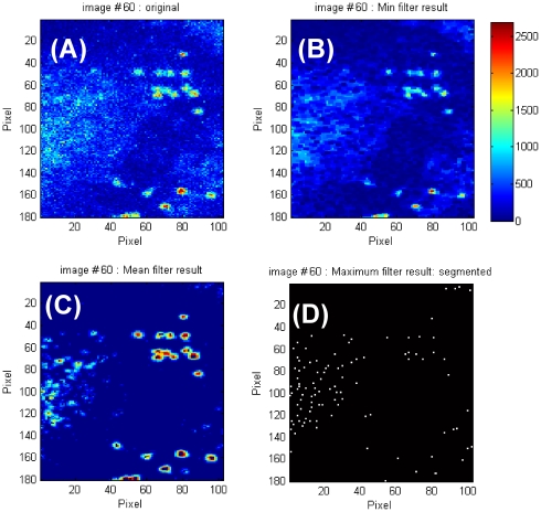
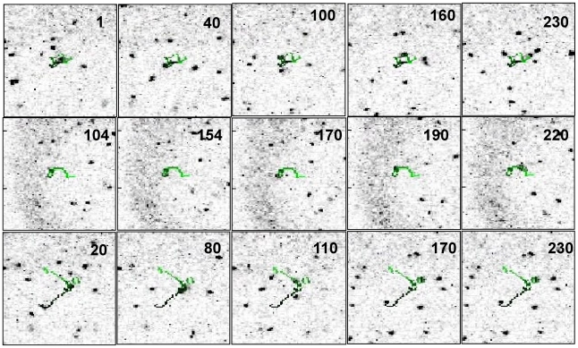
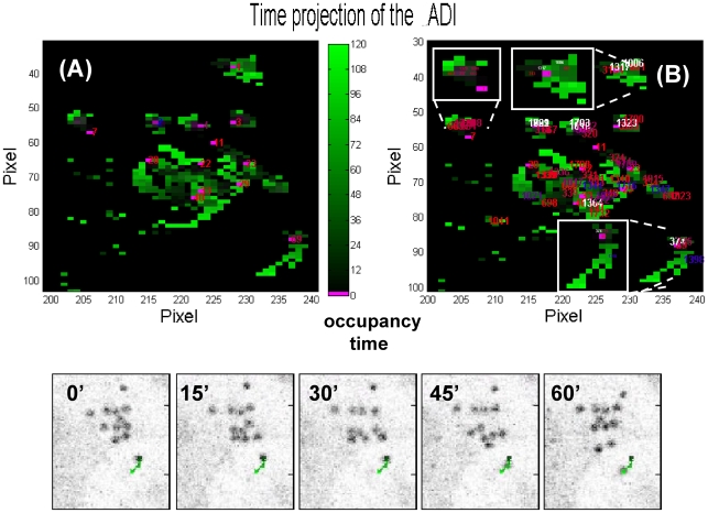
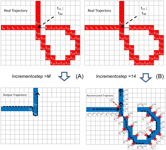
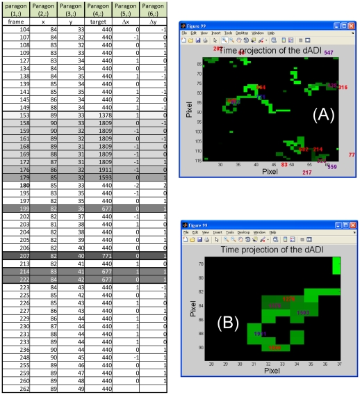
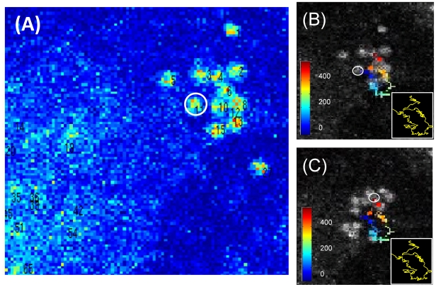
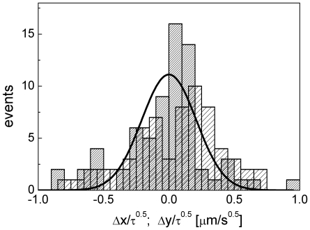
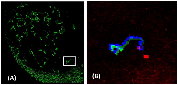
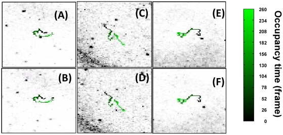
The basic research in cell biology and in medical sciences makes large use of imaging tools mainly based on confocal fluorescence and, more recently, on non-linear excitation microscopy. Substantially the aim is the recognition of selected targets in the image and their tracking in time. We have developed a particle tracking algorithm optimized for low signal/noise images with a minimum set of requirements on the target size and with no a priori knowledge of the type of motion. The image segmentation, based on a combination of size sensitive filters, does not rely on edge detection and is tailored for targets acquired at low resolution as in most of the in-vivo studies. The particle tracking is performed by building, from a stack of Accumulative Difference Images, a single 2D image in which the motion of the whole set of the particles is coded in time by a color level. This algorithm, tested here on solid-lipid nanoparticles diffusing within cells and on lymphocytes diffusing in lymphonodes, appears to be particularly useful for the cellular and the in-vivo microscopy image processing in which few a priori assumption on the type, the extent and the variability of particle motions, can be done.

摘要

细胞生物学和医学领域的基础研究大量使用基于共焦荧光的成像工具,最近还使用基于非线性激发的显微镜。其主要目的是识别图像中的选定目标并跟踪其随时间的变化。我们开发了一种粒子跟踪算法,该算法针对低信噪比图像进行了优化,对目标大小的要求最小,并且对运动类型没有先验知识。基于大小敏感滤波器的图像分割不依赖于边缘检测,并且针对在大多数体内研究中以低分辨率采集的目标进行了调整。通过从累积差分图像堆栈构建单个 2D 图像,粒子跟踪可以完成,其中整个粒子集的运动通过颜色级别在时间上进行编码。该算法已在细胞内扩散的固体脂质纳米粒子和淋巴结内扩散的淋巴细胞上进行了测试,对于细胞和体内显微镜图像处理非常有用,因为可以对粒子运动的类型、程度和可变性进行很少的先验假设。

https://cdn.ncbi.nlm.nih.gov/pmc/blobs/0417/2923183/16c5847933cf/pone.0012216.g009.jpg
https://cdn.ncbi.nlm.nih.gov/pmc/blobs/0417/2923183/bfd946421fbd/pone.0012216.g001.jpg
https://cdn.ncbi.nlm.nih.gov/pmc/blobs/0417/2923183/9cf00d8737f5/pone.0012216.g002.jpg
https://cdn.ncbi.nlm.nih.gov/pmc/blobs/0417/2923183/dc14f2eb745b/pone.0012216.g003.jpg
https://cdn.ncbi.nlm.nih.gov/pmc/blobs/0417/2923183/4bb460e89898/pone.0012216.g004.jpg
https://cdn.ncbi.nlm.nih.gov/pmc/blobs/0417/2923183/57ec6e182cc6/pone.0012216.g005.jpg
https://cdn.ncbi.nlm.nih.gov/pmc/blobs/0417/2923183/e2addb44a26c/pone.0012216.g006.jpg
https://cdn.ncbi.nlm.nih.gov/pmc/blobs/0417/2923183/31af0defc78d/pone.0012216.g007.jpg
https://cdn.ncbi.nlm.nih.gov/pmc/blobs/0417/2923183/6c8d65358be3/pone.0012216.g008.jpg
https://cdn.ncbi.nlm.nih.gov/pmc/blobs/0417/2923183/16c5847933cf/pone.0012216.g009.jpg

相似文献

1
Accumulative difference image protocol for particle tracking in fluorescence microscopy tested in mouse lymphonodes.累积差分图像协议在小鼠淋巴结的荧光显微镜粒子跟踪中的应用测试。
PLoS One. 2010 Aug 17;5(8):e12216. doi: 10.1371/journal.pone.0012216.
2
[A tracking algorithm for live mitochondria in fluorescent microscopy images].[荧光显微镜图像中活线粒体的跟踪算法]
Sheng Wu Yi Xue Gong Cheng Xue Za Zhi. 2012 Apr;29(2):352-8.
3
FocAn: automated 3D analysis of DNA repair foci in image stacks acquired by confocal fluorescence microscopy.FocAn:通过共聚焦荧光显微镜获取的图像堆栈中 DNA 修复焦点的自动 3D 分析。
BMC Bioinformatics. 2020 Jan 28;21(1):27. doi: 10.1186/s12859-020-3370-8.
4
Development of fan-shaped tracker for single particle tracking.扇型追踪器用于单颗粒追踪的研制。
Microsc Res Tech. 2020 Sep;83(9):1056-1065. doi: 10.1002/jemt.23496. Epub 2020 Apr 23.
5
A novel multiple hypothesis based particle tracking method for clathrin mediated endocytosis analysis using fluorescence microscopy.一种新型基于多重假设的粒子跟踪方法,用于使用荧光显微镜分析网格蛋白介导的胞吞作用。
IEEE Trans Image Process. 2014 Apr;23(4):1844-57. doi: 10.1109/TIP.2014.2303633.
6
Using CellX to quantify intracellular events.使用CellX对细胞内事件进行定量分析。
Curr Protoc Mol Biol. 2013;Chapter 14:Unit 14.22.. doi: 10.1002/0471142727.mb1422s101.
7
Cega: a single particle segmentation algorithm to identify moving particles in a noisy system.Cega:一种用于在噪声系统中识别运动粒子的单粒子分割算法。
Mol Biol Cell. 2021 Apr 19;32(9):931-941. doi: 10.1091/mbc.E20-11-0744. Epub 2021 Mar 31.
8
A new framework for particle detection in low-SNR fluorescence live-cell images and its application for improved particle tracking.一种用于低 SNR 荧光活细胞图像中粒子检测的新框架及其在改进粒子跟踪中的应用。
IEEE Trans Biomed Eng. 2012 Jul;59(7):2040-50. doi: 10.1109/TBME.2012.2196798. Epub 2012 Apr 27.
9
Tracking Virus Particles in Fluorescence Microscopy Images Using Multi-Scale Detection and Multi-Frame Association.使用多尺度检测和多帧关联跟踪荧光显微镜图像中的病毒颗粒。
IEEE Trans Image Process. 2015 Nov;24(11):4122-36. doi: 10.1109/TIP.2015.2458174. Epub 2015 Jul 17.
10
Pinhole Closure Improves Spatial Resolution in Confocal Scanning Microscopy.针孔闭合可提高共焦扫描显微镜的空间分辨率。
Methods Mol Biol. 2021;2274:385-389. doi: 10.1007/978-1-0716-1258-3_31.

引用本文的文献

1
Autonomous Detection of Humans in Off-Limits Mountain Areas.自主检测禁入山区的人类活动。
Sensors (Basel). 2024 Jan 25;24(3):782. doi: 10.3390/s24030782.
2
Prolonged contact with dendritic cells turns lymph node-resident NK cells into anti-tumor effectors.与树突状细胞的长时间接触会使驻留在淋巴结中的自然杀伤细胞转变为抗肿瘤效应细胞。
EMBO Mol Med. 2016 Sep 1;8(9):1039-51. doi: 10.15252/emmm.201506164. Print 2016 Sep.
3
Modeling leukocyte-leukocyte non-contact interactions in a lymph node.模拟淋巴结中白细胞与白细胞的非接触相互作用。

本文引用的文献

1
Nanoparticle image velocimetry at topologically structured surfaces.纳米粒子图像测速在拓扑结构表面上的应用。
Biomicrofluidics. 2009 Dec 1;3(4):44111. doi: 10.1063/1.3270523.
2
Automated tracking of migrating cells in phase-contrast video microscopy sequences using image registration.使用图像配准在相差视频显微镜序列中自动跟踪迁移细胞。
J Microsc. 2009 Apr;234(1):62-79. doi: 10.1111/j.1365-2818.2009.03144.x.
3
In vivo imaging and quantitative analysis of leukocyte directional migration and polarization in inflamed tissue.炎症组织中白细胞定向迁移和极化的体内成像及定量分析
PLoS One. 2013 Oct 28;8(10):e76756. doi: 10.1371/journal.pone.0076756. eCollection 2013.
4
Motion analysis of live objects by super-resolution fluorescence microscopy.利用超高分辨率荧光显微镜对活体对象进行运动分析。
Comput Math Methods Med. 2012;2012:859398. doi: 10.1155/2012/859398. Epub 2011 Nov 17.
PLoS One. 2009;4(3):e4693. doi: 10.1371/journal.pone.0004693. Epub 2009 Mar 4.
4
An automatic system for in vitro cell migration studies.
J Microsc. 2009 Jan;233(1):178-91. doi: 10.1111/j.1365-2818.2008.03108.x.
5
Two-photon autofluorescence microscopy of multicolor excitation.多色激发的双光子自发荧光显微镜术
Opt Lett. 2009 Jan 15;34(2):202-4. doi: 10.1364/ol.34.000202.
6
Multiple objects tracking in fluorescence microscopy.荧光显微镜中的多目标跟踪
J Math Biol. 2009 Jan;58(1-2):57-80. doi: 10.1007/s00285-008-0180-4. Epub 2008 May 14.
7
Dendritic cell behaviour in vivo: lessons learned from intravital two-photon microscopy.体内树突状细胞行为:从活体双光子显微镜学到的经验教训。
Immunol Cell Biol. 2008 Jul;86(5):428-38. doi: 10.1038/icb.2008.25. Epub 2008 Apr 22.
8
Image filtering for two-photon deep imaging of lymphonodes.用于淋巴结双光子深度成像的图像滤波
Eur Biophys J. 2008 Jul;37(6):979-87. doi: 10.1007/s00249-008-0323-y. Epub 2008 Apr 4.
9
Decoding the dynamics of T cell-dendritic cell interactions in vivo.解码体内T细胞与树突状细胞相互作用的动态过程。
Immunol Rev. 2008 Feb;221:182-7. doi: 10.1111/j.1600-065X.2008.00588.x.
10
Active contours without edges.无边缘活动轮廓。
IEEE Trans Image Process. 2001;10(2):266-77. doi: 10.1109/83.902291.