Department of Biomedical Engineering, The City College of New York, The City University of New York, New York, New York, United States of America.
PLoS One. 2010 Aug 16;5(8):e12196. doi: 10.1371/journal.pone.0012196.
During vascular injury, vascular smooth muscle cells (SMCs) and fibroblasts/myofibroblasts (FBs/MFBs) are exposed to altered luminal blood flow or transmural interstitial flow. We investigate the effects of these two types of fluid flows on the phenotypes of SMCs and MFBs and the underlying mechanotransduction mechanisms.
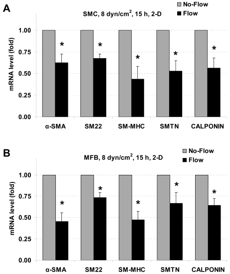
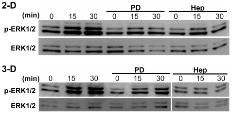
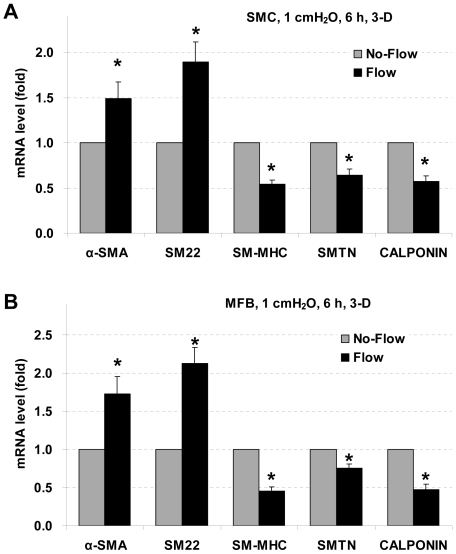
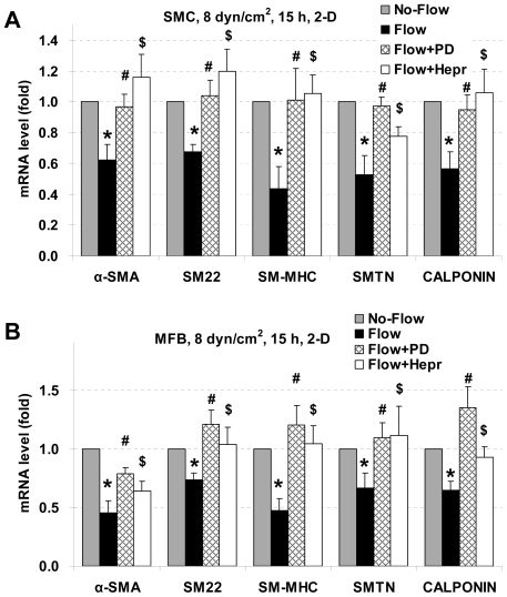
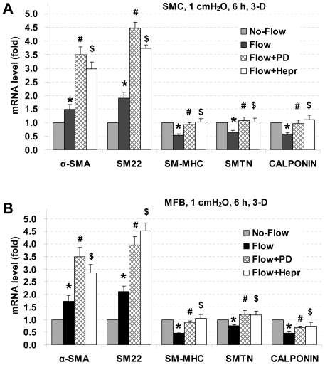
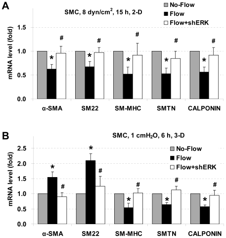
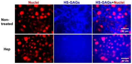
METHODOLOGY/PRINCIPAL FINDINGS: Exposure to 8 dyn/cm(2) laminar flow shear stress (2-dimensional, 2-D) for 15 h significantly reduced expression of alpha-smooth muscle actin (alpha-SMA), smooth muscle protein 22 (SM22), SM myosin heavy chain (SM-MHC), smoothelin, and calponin. Cells suspended in collagen gels were exposed to interstitial flow (1 cmH(2)O, approximately 0.05 dyn/cm(2), 3-D), and after 6 h of exposure, expression of SM-MHC, smoothelin, and calponin were significantly reduced, while expression of alpha-SMA and SM22 were markedly enhanced. PD98059 (an ERK1/2 inhibitor) and heparinase III (an enzyme to cleave heparan sulfate) significantly blocked the effects of laminar flow on gene expression, and also reversed the effects of interstitial flow on SM-MHC, smoothelin, and calponin, but enhanced interstitial flow-induced expression of alpha-SMA and SM22. SMCs and MFBs have similar responses to fluid flow. Silencing ERK1/2 completely blocked the effects of both laminar flow and interstitial flow on SMC marker gene expression. Western blotting showed that both types of flows induced ERK1/2 activation that was inhibited by disruption of heparan sulfate proteoglycans (HSPGs).
CONCLUSIONS/SIGNIFICANCE: The results suggest that HSPG-mediated ERK1/2 activation is an important mechanotransduction pathway modulating SMC marker gene expression when SMCs and MFBs are exposed to flow. Fluid flow may be involved in vascular remodeling and lesion formation by affecting phenotypes of vascular wall cells. This study has implications in understanding the flow-related mechanobiology in vascular lesion formation, tumor cell invasion, and stem cell differentiation.
在血管损伤过程中,血管平滑肌细胞(SMCs)和成纤维细胞/肌成纤维细胞(FBs/MFBs)会暴露于变化的管腔血流或跨壁间质流。我们研究了这两种类型的流体流动对 SMCs 和 MFBs 表型的影响及其潜在的力学转导机制。
方法/主要发现:SMC 暴露于 8 dyn/cm(2)层流切应力(二维,2-D)15 小时后,α-平滑肌肌动蛋白(α-SMA)、平滑肌蛋白 22(SM22)、SM 肌球蛋白重链(SM-MHC)、smoothelin 和 calponin 的表达明显降低。悬浮在胶原凝胶中的细胞暴露于间质流(1 cmH(2)O,约 0.05 dyn/cm(2),3-D),暴露 6 小时后,SM-MHC、smoothelin 和 calponin 的表达明显降低,而 α-SMA 和 SM22 的表达显著增强。PD98059(ERK1/2 抑制剂)和肝素酶 III(一种裂解肝素硫酸盐的酶)显著阻断了层流对基因表达的影响,也逆转了间质流对 SM-MHC、smoothelin 和 calponin 的影响,但增强了间质流诱导的 α-SMA 和 SM22 的表达。SMC 和 MFB 对流体流动有相似的反应。ERK1/2 的沉默完全阻断了层流和间质流对 SMC 标记基因表达的影响。Western 印迹显示,两种类型的流动都诱导了 ERK1/2 的激活,而肝素硫酸盐蛋白聚糖(HSPGs)的破坏抑制了这种激活。
结论/意义:结果表明,当 SMCs 和 MFBs 暴露于流动时,HSPG 介导的 ERK1/2 激活是调节 SMC 标记基因表达的重要力学转导途径。流体流动可能通过影响血管壁细胞的表型参与血管重塑和病变形成。本研究对理解与血流相关的血管病变形成、肿瘤细胞侵袭和干细胞分化中的力学生物学具有重要意义。