Key Laboratory of Medical Protection for Electromagnetic Radiation Ministry of Education, Third Military Medical University, Chongqing, China.
J Neuroinflammation. 2010 Sep 9;7:54. doi: 10.1186/1742-2094-7-54.
In several neuropathological conditions, microglia can become overactivated and cause neurotoxicity by initiating neuronal damage in response to pro-inflammatory stimuli. Our previous studies have shown that exposure to electromagnetic fields (EMF) activates cultured microglia to produce tumor necrosis factor (TNF)-α and nitric oxide (NO) through signal transduction involving the activator of transcription STAT3. Here, we investigated the role of STAT3 signaling in EMF-induced microglial activation and pro-inflammatory responses in more detail than the previous study.
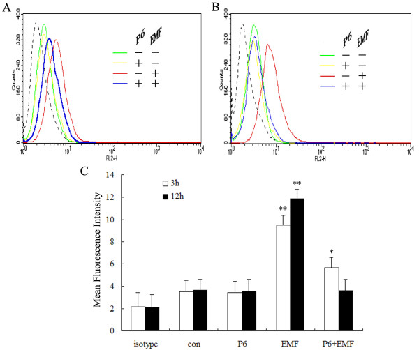
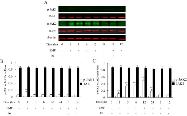
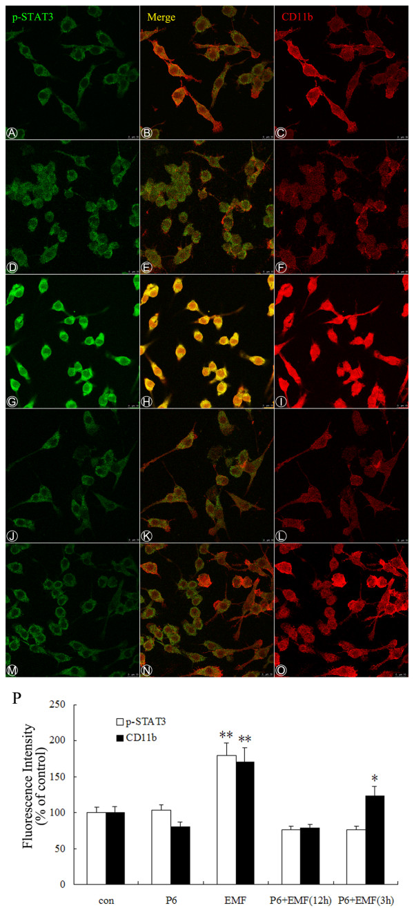
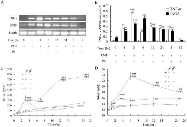
N9 microglial cells were treated with EMF exposure or a sham treatment, with or without pretreatment with an inhibitor (Pyridone 6, P6) of the Janus family of tyrosine kinases (JAK). The activation state of microglia was assessed via immunoreaction using the microglial marker CD11b. Levels of inducible nitric oxide synthase (iNOS), TNF-α and NO were measured using real-time reverse transcription-polymerase chain reaction (RT-PCR), enzyme-linked immunosorbent assay (ELISA) and the nitrate reductase method. Activation of JAKs and STAT3 proteins was evaluated by western blotting for specific tyrosine phosphorylation. The ability of STAT3 to bind to DNA was detected with an electrophoresis mobility shift assay (EMSA).
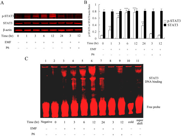
EMF was found to significantly induce phosphorylation of JAK2 and STAT3, and DNA-binding ability of STAT3 in N9 microglia. In addition, EMF dramatically increased the expression of CD11b, TNF-α and iNOS, and the production of NO. P6 strongly suppressed the phosphorylation of JAK2 and STAT3 and diminished STAT3 activity in EMF-stimulated microglia. Interestingly, expression of CD11b as well as gene expression and production of TNF-α and iNOS were suppressed by P6 at 12 h, but not at 3 h, after EMF exposure.
EMF exposure directly triggers initial activation of microglia and produces a significant pro-inflammatory response. Our findings confirm that the JAK2-STAT3 pathway may not mediate this initial microglial activation but does promote pro-inflammatory responses in EMF-stimulated microglial cells. Thus, the JAK2-STAT3 pathway might be a therapeutic target for reducing pro-inflammatory responses in EMF-activated microglia.
在几种神经病理学条件下,小胶质细胞可能会过度激活,并通过响应促炎刺激引发神经元损伤而导致神经毒性。我们之前的研究表明,暴露于电磁场 (EMF) 通过涉及转录激活因子 STAT3 的信号转导,使培养的小胶质细胞激活以产生肿瘤坏死因子 (TNF)-α 和一氧化氮 (NO)。在这里,我们比之前的研究更详细地研究了 STAT3 信号在 EMF 诱导的小胶质细胞激活和促炎反应中的作用。
用 EMF 暴露或假处理、或不用 Janus 家族酪氨酸激酶 (JAK) 抑制剂 (Pyridone 6, P6) 预处理 N9 小胶质细胞。通过使用小胶质细胞标志物 CD11b 的免疫反应评估小胶质细胞的激活状态。使用实时逆转录-聚合酶链反应 (RT-PCR)、酶联免疫吸附测定 (ELISA) 和硝酸盐还原酶法测量诱导型一氧化氮合酶 (iNOS)、TNF-α 和 NO 的水平。通过 Western 印迹评估 JAKs 和 STAT3 蛋白的激活情况,检测特定酪氨酸磷酸化。通过电泳迁移率变动分析 (EMSA) 检测 STAT3 与 DNA 结合的能力。
发现 EMF 可显著诱导 N9 小胶质细胞中 JAK2 和 STAT3 的磷酸化以及 STAT3 的 DNA 结合能力。此外,EMF 可显著增加 CD11b、TNF-α 和 iNOS 的表达以及 NO 的产生。P6 强烈抑制 EMF 刺激的小胶质细胞中 JAK2 和 STAT3 的磷酸化并降低 STAT3 活性。有趣的是,在 EMF 暴露后 12 小时而非 3 小时,P6 抑制了 CD11b 的表达以及 TNF-α 和 iNOS 的基因表达和产生。
EMF 暴露直接触发小胶质细胞的初始激活并产生显著的促炎反应。我们的发现证实,JAK2-STAT3 途径可能不会介导这种初始小胶质细胞激活,但确实促进了 EMF 刺激的小胶质细胞中的促炎反应。因此,JAK2-STAT3 途径可能是减少 EMF 激活的小胶质细胞中促炎反应的治疗靶标。